

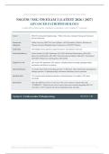
WILKES UNIVERSITY | GRADUATE NURSING AND ADVANCED PRACTICE EDUCATION
NSG530 / NSG 530 EXAM 2 (LATEST )
ADVANCED PATHOPHYSIOLOGY
COMPLETE GUIDE WITH VERIFIED ANSWERS | 100% CORRECT - WILKES
Course NSG 530 Advanced Pathophysiology - Wilkes University, Graduate Nursing and Advanced
Practice Education
Framework Wilkes University NSG 530 Course Syllabus; AACN Essentials of Master's Education in
Alignment Nursing; Advanced Pathophysiology Competencies (2026/2027 Edition)
Total Items 100 multiple-choice questions, single best answer, four options (A-D) each
Section Breakdown Cardiovascular (Q1-Q20); Pulmonary (Q21-Q35); Renal and Genitourinary (Q36-Q50);
Endocrine and Metabolic (Q51-Q65); Gastrointestinal and Hepatic (Q66-Q77); Neurological
(Q78-Q90); Multisystem and Integrative (Q91-Q100)
Cognitive Levels 20% recall; 50% application; 30% analysis, including clinical reasoning, pathophysiologic
mechanisms and clinical correlations
Special Inclusions 15 scenario-based clinical reasoning questions; 10 laboratory value interpretation and diagnostic
findings questions; 10 pathophysiologic mechanism and disease process questions
Question Style 75% scenario-based (patient presentations, clinical situations, case studies); 25% direct
knowledge
Instructions Select the ONE best answer for each item. The correct option is marked inline with
*[CORRECT]*, followed by the correct answer letter and a rationale integrating NSG 530
curriculum, AACN standards and advanced pathophysiology principles.
Section 1: Cardiovascular Pathophysiology Questions 1-20
Wilkes University | Graduate Nursing | NSG 530 Advanced Pathophysiology 1
,NSG530 / NSG 530 EXAM 2 () - ADVANCED PATHOPHYSIOLOGY COMPLETE GUIDE WITH VERIFIED ANSWERS
Q1. Which sequence best describes the earliest pathophysiologic events in the formation of an atherosclerotic
plaque?
A. Transmural myocardial inflammation followed by necrotic core formation and aneurysm dilation
B. Endothelial injury and dysfunction allowing low-density lipoprotein entry into the intima, LDL
oxidation, monocyte recruitment and macrophage foam cell formation *[CORRECT]*
C. Rupture of a thin-cap fibroatheroma with acute platelet-rich thrombus formation
D. Medial calcification and stiffening of muscular arteries with loss of elastic recoil
Correct Answer: B
Rationale: Atherogenesis begins with endothelial dysfunction produced by hypertension, smoking, hyperlipidemia
and diabetes, which permits LDL to enter and be retained in the intima. Oxidized LDL recruits monocytes that
become macrophages and engulf lipid to form foam cells, generating the fatty streak, the earliest visible lesion
(NSG 530 vascular pathology). Choice C describes rupture of an established unstable plaque, a late event
responsible for acute coronary syndromes, while choice D describes Monckeberg medial calcific sclerosis, which
does not narrow the lumen.
Q2. A 62-year-old man reports substernal pressure after walking two blocks that resolves within minutes of
resting and responds to sublingual nitroglycerin. ECG during pain shows transient ST-segment depression.
Which mechanism best explains his chest pain?
A. Transient platelet-rich thrombus completely occluding a major epicardial coronary artery
B. Coronary vasospasm occurring at rest with transient ST-segment elevation
C. Fixed atherosclerotic stenosis limiting coronary flow reserve, so exertional increases in myocardial
oxygen demand produce reversible subendocardial ischemia *[CORRECT]*
D. Spontaneous coronary artery dissection reducing antegrade flow in the left main artery
Correct Answer: C
Rationale: Stable angina reflects supply-demand mismatch across a fixed atherosclerotic narrowing; exertion
raises myocardial oxygen demand beyond stenosis-limited supply, producing subendocardial ischemia with ST
depression that reverses with rest. Complete thrombotic occlusion (choice A) defines acute coronary syndrome
with myocardial injury, while rest-associated vasospasm with ST elevation (choice B) defines Prinzmetal variant
angina. Spontaneous dissection (choice D) typically produces infarction in younger and peripartum patients and
does not resolve with rest.
Q3. A 58-year-old woman presents with crushing substernal chest pain for 3 hours and ST elevation in leads II,
III and aVF. Which laboratory pattern confirms the diagnosis of acute myocardial infarction?
A. Elevated high-sensitivity cardiac troponin I with a rising delta value on serial testing, indicating ongoing
myocyte necrosis *[CORRECT]*
B. Elevated CK-MB with a completely normal troponin level 6 hours after symptom onset
C. Isolated lactate dehydrogenase elevation with normal cardiac troponin
D. A single normal troponin at presentation that permanently excludes myocardial infarction
Correct Answer: A
Rationale: Cardiac troponin I and T are the most specific and sensitive markers of myocyte injury, rising within 2
to 4 hours, peaking at 18 to 24 hours and remaining elevated for 7 to 10 days; a rising serial delta confirms acute
injury. A single normal value at presentation never excludes infarction because levels may not yet be detectable,
which is why serial sampling is required (NSG 530 diagnostic reasoning). LDH is obsolete for cardiac diagnosis,
and CK-MB alone with normal troponin does not confirm acute myocardial necrosis.
Wilkes University | Graduate Nursing | NSG 530 Advanced Pathophysiology 2
,NSG530 / NSG 530 EXAM 2 () - ADVANCED PATHOPHYSIOLOGY COMPLETE GUIDE WITH VERIFIED ANSWERS
Q4. Five days after an inferior myocardial infarction, a 67-year-old man develops acute pulmonary edema and
cardiogenic shock. Auscultation reveals a new holosystolic murmur at the apex radiating to the axilla. Which
complication best explains this presentation?
A. Ventricular free wall rupture with cardiac tamponade
B. Rheumatic mitral stenosis from commissural fusion
C. Aortic dissection extending into the aortic root
D. Papillary muscle rupture producing acute mitral regurgitation *[CORRECT]*
Correct Answer: D
Rationale: Papillary muscle rupture typically occurs 2 to 7 days post-infarction, classically involving the
posteromedial papillary muscle, which has a single blood supply from the posterior descending artery. The loss of
chordal support produces acute severe mitral regurgitation with a holosystolic apical murmur and flash pulmonary
edema. Free wall rupture (choice A) causes hemopericardium and tamponade without a new murmur, while
rheumatic disease (choice B) and dissection (choice C) are not acute post-MI complications.
Q5. Which statement best characterizes the pathogenesis of primary (essential) hypertension?
A. Unilateral renal artery stenosis chronically activating the renin-angiotensin-aldosterone system
B. Combined genetic and environmental factors that impair renal sodium excretion, increase sympathetic
nervous system tone and heighten vascular smooth muscle reactivity *[CORRECT]*
C. Catecholamine excess from an adrenal medullary tumor driving episodic vasoconstriction
D. Autonomous aldosterone secretion from an adrenal adenoma causing sodium retention and potassium
wasting
Correct Answer: B
Rationale: Essential hypertension is a multifactorial disorder in which inherited susceptibility interacts with
environmental factors such as obesity, high sodium intake, alcohol and stress, producing defective renal natriuresis,
sympathetic overactivity and increased vascular reactivity (NSG 530 cardiovascular regulation). Choices A, C and
D describe renal artery stenosis, pheochromocytoma and primary hyperaldosteronism, which are well-defined
secondary causes of hypertension and are common distractors on examinations.
Q6. A 47-year-old woman develops abrupt-onset, treatment-resistant hypertension. Examination reveals an
abdominal bruit and recurrent episodes of flash pulmonary edema, and her creatinine rises after starting an
ACE inhibitor. Which mechanism best explains these findings?
A. Primary hyperaldosteronism with sodium retention and volume expansion
B. Essential hypertension complicated by medication nonadherence
C. Renal artery stenosis reducing renal perfusion pressure, activating renin release, angiotensin II-mediated
vasoconstriction and aldosterone-driven volume retention, while ACE inhibition removes efferent
arteriolar tone and drops glomerular filtration *[CORRECT]*
D. Pheochromocytoma producing paroxysmal alpha-adrenergic vasoconstriction
Correct Answer: C
Rationale: Renal artery stenosis lowers perfusion pressure sensed by the juxtaglomerular apparatus, triggering
renin release, angiotensin II vasoconstriction and aldosterone-mediated volume expansion, which explains resistant
hypertension and flash pulmonary edema. Glomerular filtration in the stenotic kidney depends on angiotensin
II-mediated efferent arteriolar constriction, so ACE inhibitor or ARB therapy precipitates a rise in creatinine, a
hallmark clue. Pheochromocytoma (choice D) produces episodic headache, sweating and palpitations rather than an
abdominal bruit or ACE inhibitor-related renal decline.
Wilkes University | Graduate Nursing | NSG 530 Advanced Pathophysiology 3
, NSG530 / NSG 530 EXAM 2 () - ADVANCED PATHOPHYSIOLOGY COMPLETE GUIDE WITH VERIFIED ANSWERS
Q7. A 66-year-old man has orthopnea, paroxysmal nocturnal dyspnea, a laterally displaced point of maximal
impulse and an S3 gallop. Echocardiography shows a left ventricular ejection fraction of 28 percent with
four-chamber dilation. Which mechanism primarily produces his heart failure?
A. Loss of functioning myocytes with impaired contractility, producing ventricular dilation and reduced
ejection fraction *[CORRECT]*
B. Concentric left ventricular hypertrophy with a stiff, noncompliant ventricle and preserved ejection
fraction
C. Constrictive pericarditis restricting diastolic filling of both ventricles equally
D. Mitral stenosis mechanically limiting left ventricular preload
Correct Answer: A
Rationale: Heart failure with reduced ejection fraction (HFrEF, systolic failure) results from impaired myocardial
contractility due to ischemic injury, dilated cardiomyopathy or volume overload, producing ventricular dilation, an
S3 gallop and reduced ejection fraction below 40 percent. Choice B describes heart failure with preserved ejection
fraction (HFpEF, diastolic failure), while constrictive pericarditis and mitral stenosis are distinct restriction and
inflow-obstruction states that do not primarily reduce contractility.
Q8. A 74-year-old woman with long-standing hypertension develops progressive exertional dyspnea.
Echocardiography shows an ejection fraction of 60 percent, concentric left ventricular hypertrophy and an
elevated early filling transmitral pressure ratio (E/e prime). Which mechanism best explains her pulmonary
congestion?
A. Reduced contractile force from extensive myocyte loss
B. Progressive ventricular dilation with mitral annular stretch
C. Primary renal sodium retention with excess circulating volume
D. Decreased ventricular compliance from hypertrophy and fibrosis, elevating diastolic filling pressures
despite preserved systolic function *[CORRECT]*
Correct Answer: D
Rationale: This is heart failure with preserved ejection fraction (HFpEF, diastolic dysfunction), which accounts
for roughly half of heart failure cases and is most common in older hypertensive women. Chronic pressure overload
drives concentric hypertrophy and interstitial fibrosis, stiffening the ventricle so that normal filling volumes
generate elevated left ventricular end-diastolic pressures that back up into the pulmonary circulation. Choices A
and B describe systolic failure, and choice C describes renal volume overload, none of which matches the preserved
ejection fraction with abnormal filling pressures.
Q9. Which cluster of clinical manifestations is most characteristic of right-sided heart failure?
A. Orthopnea, bilateral basilar crackles and an S3 gallop
B. Jugular venous distension, dependent peripheral edema, hepatomegaly and ascites *[CORRECT]*
C. Paroxysmal nocturnal dyspnea with pink, frothy sputum
D. Exertional dyspnea with pulmonary vascular congestion on chest radiograph
Correct Answer: B
Rationale: Right-sided failure produces systemic venous congestion because the failing right ventricle cannot
accommodate venous return, causing jugular venous distension, dependent edema, hepatomegaly, splenomegaly
and ascites. Choices A, C and D reflect pulmonary venous congestion from left-sided failure, the most common
cause of secondary right ventricular failure. Cor pulmonale is the term for right heart failure caused specifically by
primary pulmonary disease such as COPD.
Wilkes University | Graduate Nursing | NSG 530 Advanced Pathophysiology 4
NSG530 / NSG 530 EXAM 2 (LATEST )
ADVANCED PATHOPHYSIOLOGY
COMPLETE GUIDE WITH VERIFIED ANSWERS | 100% CORRECT - WILKES
Course NSG 530 Advanced Pathophysiology - Wilkes University, Graduate Nursing and Advanced
Practice Education
Framework Wilkes University NSG 530 Course Syllabus; AACN Essentials of Master's Education in
Alignment Nursing; Advanced Pathophysiology Competencies (2026/2027 Edition)
Total Items 100 multiple-choice questions, single best answer, four options (A-D) each
Section Breakdown Cardiovascular (Q1-Q20); Pulmonary (Q21-Q35); Renal and Genitourinary (Q36-Q50);
Endocrine and Metabolic (Q51-Q65); Gastrointestinal and Hepatic (Q66-Q77); Neurological
(Q78-Q90); Multisystem and Integrative (Q91-Q100)
Cognitive Levels 20% recall; 50% application; 30% analysis, including clinical reasoning, pathophysiologic
mechanisms and clinical correlations
Special Inclusions 15 scenario-based clinical reasoning questions; 10 laboratory value interpretation and diagnostic
findings questions; 10 pathophysiologic mechanism and disease process questions
Question Style 75% scenario-based (patient presentations, clinical situations, case studies); 25% direct
knowledge
Instructions Select the ONE best answer for each item. The correct option is marked inline with
*[CORRECT]*, followed by the correct answer letter and a rationale integrating NSG 530
curriculum, AACN standards and advanced pathophysiology principles.
Section 1: Cardiovascular Pathophysiology Questions 1-20
Wilkes University | Graduate Nursing | NSG 530 Advanced Pathophysiology 1
,NSG530 / NSG 530 EXAM 2 () - ADVANCED PATHOPHYSIOLOGY COMPLETE GUIDE WITH VERIFIED ANSWERS
Q1. Which sequence best describes the earliest pathophysiologic events in the formation of an atherosclerotic
plaque?
A. Transmural myocardial inflammation followed by necrotic core formation and aneurysm dilation
B. Endothelial injury and dysfunction allowing low-density lipoprotein entry into the intima, LDL
oxidation, monocyte recruitment and macrophage foam cell formation *[CORRECT]*
C. Rupture of a thin-cap fibroatheroma with acute platelet-rich thrombus formation
D. Medial calcification and stiffening of muscular arteries with loss of elastic recoil
Correct Answer: B
Rationale: Atherogenesis begins with endothelial dysfunction produced by hypertension, smoking, hyperlipidemia
and diabetes, which permits LDL to enter and be retained in the intima. Oxidized LDL recruits monocytes that
become macrophages and engulf lipid to form foam cells, generating the fatty streak, the earliest visible lesion
(NSG 530 vascular pathology). Choice C describes rupture of an established unstable plaque, a late event
responsible for acute coronary syndromes, while choice D describes Monckeberg medial calcific sclerosis, which
does not narrow the lumen.
Q2. A 62-year-old man reports substernal pressure after walking two blocks that resolves within minutes of
resting and responds to sublingual nitroglycerin. ECG during pain shows transient ST-segment depression.
Which mechanism best explains his chest pain?
A. Transient platelet-rich thrombus completely occluding a major epicardial coronary artery
B. Coronary vasospasm occurring at rest with transient ST-segment elevation
C. Fixed atherosclerotic stenosis limiting coronary flow reserve, so exertional increases in myocardial
oxygen demand produce reversible subendocardial ischemia *[CORRECT]*
D. Spontaneous coronary artery dissection reducing antegrade flow in the left main artery
Correct Answer: C
Rationale: Stable angina reflects supply-demand mismatch across a fixed atherosclerotic narrowing; exertion
raises myocardial oxygen demand beyond stenosis-limited supply, producing subendocardial ischemia with ST
depression that reverses with rest. Complete thrombotic occlusion (choice A) defines acute coronary syndrome
with myocardial injury, while rest-associated vasospasm with ST elevation (choice B) defines Prinzmetal variant
angina. Spontaneous dissection (choice D) typically produces infarction in younger and peripartum patients and
does not resolve with rest.
Q3. A 58-year-old woman presents with crushing substernal chest pain for 3 hours and ST elevation in leads II,
III and aVF. Which laboratory pattern confirms the diagnosis of acute myocardial infarction?
A. Elevated high-sensitivity cardiac troponin I with a rising delta value on serial testing, indicating ongoing
myocyte necrosis *[CORRECT]*
B. Elevated CK-MB with a completely normal troponin level 6 hours after symptom onset
C. Isolated lactate dehydrogenase elevation with normal cardiac troponin
D. A single normal troponin at presentation that permanently excludes myocardial infarction
Correct Answer: A
Rationale: Cardiac troponin I and T are the most specific and sensitive markers of myocyte injury, rising within 2
to 4 hours, peaking at 18 to 24 hours and remaining elevated for 7 to 10 days; a rising serial delta confirms acute
injury. A single normal value at presentation never excludes infarction because levels may not yet be detectable,
which is why serial sampling is required (NSG 530 diagnostic reasoning). LDH is obsolete for cardiac diagnosis,
and CK-MB alone with normal troponin does not confirm acute myocardial necrosis.
Wilkes University | Graduate Nursing | NSG 530 Advanced Pathophysiology 2
,NSG530 / NSG 530 EXAM 2 () - ADVANCED PATHOPHYSIOLOGY COMPLETE GUIDE WITH VERIFIED ANSWERS
Q4. Five days after an inferior myocardial infarction, a 67-year-old man develops acute pulmonary edema and
cardiogenic shock. Auscultation reveals a new holosystolic murmur at the apex radiating to the axilla. Which
complication best explains this presentation?
A. Ventricular free wall rupture with cardiac tamponade
B. Rheumatic mitral stenosis from commissural fusion
C. Aortic dissection extending into the aortic root
D. Papillary muscle rupture producing acute mitral regurgitation *[CORRECT]*
Correct Answer: D
Rationale: Papillary muscle rupture typically occurs 2 to 7 days post-infarction, classically involving the
posteromedial papillary muscle, which has a single blood supply from the posterior descending artery. The loss of
chordal support produces acute severe mitral regurgitation with a holosystolic apical murmur and flash pulmonary
edema. Free wall rupture (choice A) causes hemopericardium and tamponade without a new murmur, while
rheumatic disease (choice B) and dissection (choice C) are not acute post-MI complications.
Q5. Which statement best characterizes the pathogenesis of primary (essential) hypertension?
A. Unilateral renal artery stenosis chronically activating the renin-angiotensin-aldosterone system
B. Combined genetic and environmental factors that impair renal sodium excretion, increase sympathetic
nervous system tone and heighten vascular smooth muscle reactivity *[CORRECT]*
C. Catecholamine excess from an adrenal medullary tumor driving episodic vasoconstriction
D. Autonomous aldosterone secretion from an adrenal adenoma causing sodium retention and potassium
wasting
Correct Answer: B
Rationale: Essential hypertension is a multifactorial disorder in which inherited susceptibility interacts with
environmental factors such as obesity, high sodium intake, alcohol and stress, producing defective renal natriuresis,
sympathetic overactivity and increased vascular reactivity (NSG 530 cardiovascular regulation). Choices A, C and
D describe renal artery stenosis, pheochromocytoma and primary hyperaldosteronism, which are well-defined
secondary causes of hypertension and are common distractors on examinations.
Q6. A 47-year-old woman develops abrupt-onset, treatment-resistant hypertension. Examination reveals an
abdominal bruit and recurrent episodes of flash pulmonary edema, and her creatinine rises after starting an
ACE inhibitor. Which mechanism best explains these findings?
A. Primary hyperaldosteronism with sodium retention and volume expansion
B. Essential hypertension complicated by medication nonadherence
C. Renal artery stenosis reducing renal perfusion pressure, activating renin release, angiotensin II-mediated
vasoconstriction and aldosterone-driven volume retention, while ACE inhibition removes efferent
arteriolar tone and drops glomerular filtration *[CORRECT]*
D. Pheochromocytoma producing paroxysmal alpha-adrenergic vasoconstriction
Correct Answer: C
Rationale: Renal artery stenosis lowers perfusion pressure sensed by the juxtaglomerular apparatus, triggering
renin release, angiotensin II vasoconstriction and aldosterone-mediated volume expansion, which explains resistant
hypertension and flash pulmonary edema. Glomerular filtration in the stenotic kidney depends on angiotensin
II-mediated efferent arteriolar constriction, so ACE inhibitor or ARB therapy precipitates a rise in creatinine, a
hallmark clue. Pheochromocytoma (choice D) produces episodic headache, sweating and palpitations rather than an
abdominal bruit or ACE inhibitor-related renal decline.
Wilkes University | Graduate Nursing | NSG 530 Advanced Pathophysiology 3
, NSG530 / NSG 530 EXAM 2 () - ADVANCED PATHOPHYSIOLOGY COMPLETE GUIDE WITH VERIFIED ANSWERS
Q7. A 66-year-old man has orthopnea, paroxysmal nocturnal dyspnea, a laterally displaced point of maximal
impulse and an S3 gallop. Echocardiography shows a left ventricular ejection fraction of 28 percent with
four-chamber dilation. Which mechanism primarily produces his heart failure?
A. Loss of functioning myocytes with impaired contractility, producing ventricular dilation and reduced
ejection fraction *[CORRECT]*
B. Concentric left ventricular hypertrophy with a stiff, noncompliant ventricle and preserved ejection
fraction
C. Constrictive pericarditis restricting diastolic filling of both ventricles equally
D. Mitral stenosis mechanically limiting left ventricular preload
Correct Answer: A
Rationale: Heart failure with reduced ejection fraction (HFrEF, systolic failure) results from impaired myocardial
contractility due to ischemic injury, dilated cardiomyopathy or volume overload, producing ventricular dilation, an
S3 gallop and reduced ejection fraction below 40 percent. Choice B describes heart failure with preserved ejection
fraction (HFpEF, diastolic failure), while constrictive pericarditis and mitral stenosis are distinct restriction and
inflow-obstruction states that do not primarily reduce contractility.
Q8. A 74-year-old woman with long-standing hypertension develops progressive exertional dyspnea.
Echocardiography shows an ejection fraction of 60 percent, concentric left ventricular hypertrophy and an
elevated early filling transmitral pressure ratio (E/e prime). Which mechanism best explains her pulmonary
congestion?
A. Reduced contractile force from extensive myocyte loss
B. Progressive ventricular dilation with mitral annular stretch
C. Primary renal sodium retention with excess circulating volume
D. Decreased ventricular compliance from hypertrophy and fibrosis, elevating diastolic filling pressures
despite preserved systolic function *[CORRECT]*
Correct Answer: D
Rationale: This is heart failure with preserved ejection fraction (HFpEF, diastolic dysfunction), which accounts
for roughly half of heart failure cases and is most common in older hypertensive women. Chronic pressure overload
drives concentric hypertrophy and interstitial fibrosis, stiffening the ventricle so that normal filling volumes
generate elevated left ventricular end-diastolic pressures that back up into the pulmonary circulation. Choices A
and B describe systolic failure, and choice C describes renal volume overload, none of which matches the preserved
ejection fraction with abnormal filling pressures.
Q9. Which cluster of clinical manifestations is most characteristic of right-sided heart failure?
A. Orthopnea, bilateral basilar crackles and an S3 gallop
B. Jugular venous distension, dependent peripheral edema, hepatomegaly and ascites *[CORRECT]*
C. Paroxysmal nocturnal dyspnea with pink, frothy sputum
D. Exertional dyspnea with pulmonary vascular congestion on chest radiograph
Correct Answer: B
Rationale: Right-sided failure produces systemic venous congestion because the failing right ventricle cannot
accommodate venous return, causing jugular venous distension, dependent edema, hepatomegaly, splenomegaly
and ascites. Choices A, C and D reflect pulmonary venous congestion from left-sided failure, the most common
cause of secondary right ventricular failure. Cor pulmonale is the term for right heart failure caused specifically by
primary pulmonary disease such as COPD.
Wilkes University | Graduate Nursing | NSG 530 Advanced Pathophysiology 4

