

1
610 Study Guide #4
Kidney
- Azotemia, BUN: Cr ratio
● Azotemia: A laboratory diagnosis of elevated BUN & Cr
● Uremia: Is azotemia associated with direct clinical manifestations of renal failure, such as:
○ Pericarditis
○ Increased tendency to bleeding (defective platelet function)
○ Increased liability to infections (defective WBC production)
BUN:CR ratio
● Normally, 15:1
● In renal failure, both BUN & Cr will increased because of the decreased GFR
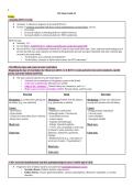
● We use BUN:Cr ratio to differentiate between the 3 types of acute renal failure (pre-, intra-, and post-renal) making use of
the fact that both urea and creatinine are filtered at the glomerulus but only urea gets reabsorbed, and only creatinine gets
secreted at the renal tubules
○ “In the kidney, urea is filtered and reabsorbed.”
○ “In the kidney, creatinine is filtered and secreted, but NOT reabsorbed.”
- The different types and causes of acute renal failure
-Diagnosing the type of renal failure by: Blood tests (BUN, Cr & BUN:Cr ratio) and urine tests (urine osmolarity, specific
gravity, and urine sodium and FENa)
● The most common indicator of acute renal failure is:
○ Azotemia (an accumulation of nitrogenous wastes in the blood)
○ A decrease in glomerular filtration rate (GFR)
○ Clues to the “acuteness”: Normal kidney size, hematocrit, and Ca++
○ Major causes of acute renal failure (ARF): Prerenal, Renal, and Post-renal
Pre-renal Renal Post-renal
Mechanism: Low blood flow getting into Mechanism: Problem with the nephron Mechanism: Urinary tract obstruction
the kidney (e.g., low perfusion) (e.g., glomerulus or tubules) causing back pressure into the kidney
Causes: Causes: Causes:
- Heart failure - Nephrotoxic medications - Benign prostatic hyperplasia
- Hypovolemia (Aminoglycosides, Vancomycin) - Cervical cancer
- Constrictive pericarditis (liver cirrhosis) - Acute tubular necrosis - Stones (bladder)
- Renal artery stenosis - Contrast agents - Neurogenic bladder
- Cisplatin (chemotherapy)
Blood tests: - Allergic interstitial nephritis Blood tests:
>> Fever, rash, eosinophils in urine stain
by Hansel’s stain), penicillin, sulfa,
Urine tests: phenytoin, allopurinol, quinolones, Urine tests:
rifampin
- Rhabdomyolysis (e.g., crush injury or
recent use of statins > hyperkalemia!)
Blood tests:
Urine tests:
- CKD: associated manifestations and their pathophysiological causes. Earliest sign of CKD.
● Progressive loss of kidney function associated with (pathophysiological causes):
○ Systemic disease: Diabetes mellitus, Hypertension
○ Kidney disease: Chronic glomerulonephritis, Chronic pyelonephritis, Obstructive uropathies, or vascular
disorders
, 2
● Clinical manifestations:
○ CKD is defined as GFR < 60 mL/min/1.73m2 for 3 months or more, irrespective of the cause
○ Initially, no symptoms (only laboratory findings!)
■ Earliest sign: Microalbuminuria, early bone disease, BUN & Cr start to increase
○ Uremia
■ Lethargy
■ Pericarditis
■ Encephalitis
■ Bleeding diathesis
■ Recurrent infections
○ Fluid volume overload
○ Hyper: -kalemia, magnesemia, phosphatemia, and metabolic acidosis
○ Hypocalcemia
○ Osteodystrophy
○ Anemia: Lack of EPO
Neurological Disorders
- Neurologic disorders: congenital, traumatic, vascular, infectious, degenerative
Congenital
● Neural tube defects
○ Anencephaly
■ The soft, bony component of the skull and part of the brain are missing
■ Results in spontaneous abortion or early neonatal death
○ Spina bifida occulta
■ Failure of fusion of the posterior vertebral laminae. Symptoms due to tethering of spinal cord (gait
abnormalities, foot deformities, and bladder sphincter disturbance)
○ Miningocele
■ A sac-like cyst of meninges filled with spinal fluid, protruding through the vertebral defect
○ Meningomyelocele (spina bifida cystica)
■ Hernial protrusion of a meningocele containing a portion of the spinal cord w/ its nerves (worst one!)
○ Encephalocele
■ Herniation of the brain and meninges through a defect in the skull
● Congenital hydrocephalus
○ Characterized by enlargement of the cerebral ventricles due to blockage of where CSF flows
○ Pushes and compresses the brain tissue against the skull cavity
○ If this develops before fusion of the cranial sutures, the skull can expand to accommodate this additional volume
● Cerebral palsy
○ A group of nonprogressive syndromes that affect the brain and cause motor dysfunction beginning in early
infancy
○ Causes
■ Prenatal or perinatal cerebral hypoxia, hemorrhage, or infection
○ Types
■ Spastic (70-80%): Hyperactive deep tendon reflexes, clonus, rigidity
■ Dystonic (10-20%): Difficulty in fine motor coordination, stiff, abrupt movements due to basal ganglia injury
■ Ataxic (5-10%): Manifests with gait disturbance and instability
Traumatic
● Closed (blunt)/open trauma
● Coup/Countercoup injury
● Focal brain injury
○ ⅔ of head injury deaths
○ Example: Extradural hematoma; Subdural hematoma
■ Extradural hematoma
● Majority: Aterial
● Lucid interval
■ Subdural hematoma
● Majority: Venous
610 Study Guide #4
Kidney
- Azotemia, BUN: Cr ratio
● Azotemia: A laboratory diagnosis of elevated BUN & Cr
● Uremia: Is azotemia associated with direct clinical manifestations of renal failure, such as:
○ Pericarditis
○ Increased tendency to bleeding (defective platelet function)
○ Increased liability to infections (defective WBC production)
BUN:CR ratio
● Normally, 15:1
● In renal failure, both BUN & Cr will increased because of the decreased GFR
● We use BUN:Cr ratio to differentiate between the 3 types of acute renal failure (pre-, intra-, and post-renal) making use of
the fact that both urea and creatinine are filtered at the glomerulus but only urea gets reabsorbed, and only creatinine gets
secreted at the renal tubules
○ “In the kidney, urea is filtered and reabsorbed.”
○ “In the kidney, creatinine is filtered and secreted, but NOT reabsorbed.”
- The different types and causes of acute renal failure
-Diagnosing the type of renal failure by: Blood tests (BUN, Cr & BUN:Cr ratio) and urine tests (urine osmolarity, specific
gravity, and urine sodium and FENa)
● The most common indicator of acute renal failure is:
○ Azotemia (an accumulation of nitrogenous wastes in the blood)
○ A decrease in glomerular filtration rate (GFR)
○ Clues to the “acuteness”: Normal kidney size, hematocrit, and Ca++
○ Major causes of acute renal failure (ARF): Prerenal, Renal, and Post-renal
Pre-renal Renal Post-renal
Mechanism: Low blood flow getting into Mechanism: Problem with the nephron Mechanism: Urinary tract obstruction
the kidney (e.g., low perfusion) (e.g., glomerulus or tubules) causing back pressure into the kidney
Causes: Causes: Causes:
- Heart failure - Nephrotoxic medications - Benign prostatic hyperplasia
- Hypovolemia (Aminoglycosides, Vancomycin) - Cervical cancer
- Constrictive pericarditis (liver cirrhosis) - Acute tubular necrosis - Stones (bladder)
- Renal artery stenosis - Contrast agents - Neurogenic bladder
- Cisplatin (chemotherapy)
Blood tests: - Allergic interstitial nephritis Blood tests:
>> Fever, rash, eosinophils in urine stain
by Hansel’s stain), penicillin, sulfa,
Urine tests: phenytoin, allopurinol, quinolones, Urine tests:
rifampin
- Rhabdomyolysis (e.g., crush injury or
recent use of statins > hyperkalemia!)
Blood tests:
Urine tests:
- CKD: associated manifestations and their pathophysiological causes. Earliest sign of CKD.
● Progressive loss of kidney function associated with (pathophysiological causes):
○ Systemic disease: Diabetes mellitus, Hypertension
○ Kidney disease: Chronic glomerulonephritis, Chronic pyelonephritis, Obstructive uropathies, or vascular
disorders
, 2
● Clinical manifestations:
○ CKD is defined as GFR < 60 mL/min/1.73m2 for 3 months or more, irrespective of the cause
○ Initially, no symptoms (only laboratory findings!)
■ Earliest sign: Microalbuminuria, early bone disease, BUN & Cr start to increase
○ Uremia
■ Lethargy
■ Pericarditis
■ Encephalitis
■ Bleeding diathesis
■ Recurrent infections
○ Fluid volume overload
○ Hyper: -kalemia, magnesemia, phosphatemia, and metabolic acidosis
○ Hypocalcemia
○ Osteodystrophy
○ Anemia: Lack of EPO
Neurological Disorders
- Neurologic disorders: congenital, traumatic, vascular, infectious, degenerative
Congenital
● Neural tube defects
○ Anencephaly
■ The soft, bony component of the skull and part of the brain are missing
■ Results in spontaneous abortion or early neonatal death
○ Spina bifida occulta
■ Failure of fusion of the posterior vertebral laminae. Symptoms due to tethering of spinal cord (gait
abnormalities, foot deformities, and bladder sphincter disturbance)
○ Miningocele
■ A sac-like cyst of meninges filled with spinal fluid, protruding through the vertebral defect
○ Meningomyelocele (spina bifida cystica)
■ Hernial protrusion of a meningocele containing a portion of the spinal cord w/ its nerves (worst one!)
○ Encephalocele
■ Herniation of the brain and meninges through a defect in the skull
● Congenital hydrocephalus
○ Characterized by enlargement of the cerebral ventricles due to blockage of where CSF flows
○ Pushes and compresses the brain tissue against the skull cavity
○ If this develops before fusion of the cranial sutures, the skull can expand to accommodate this additional volume
● Cerebral palsy
○ A group of nonprogressive syndromes that affect the brain and cause motor dysfunction beginning in early
infancy
○ Causes
■ Prenatal or perinatal cerebral hypoxia, hemorrhage, or infection
○ Types
■ Spastic (70-80%): Hyperactive deep tendon reflexes, clonus, rigidity
■ Dystonic (10-20%): Difficulty in fine motor coordination, stiff, abrupt movements due to basal ganglia injury
■ Ataxic (5-10%): Manifests with gait disturbance and instability
Traumatic
● Closed (blunt)/open trauma
● Coup/Countercoup injury
● Focal brain injury
○ ⅔ of head injury deaths
○ Example: Extradural hematoma; Subdural hematoma
■ Extradural hematoma
● Majority: Aterial
● Lucid interval
■ Subdural hematoma
● Majority: Venous

