Acute posterior eye presentations.
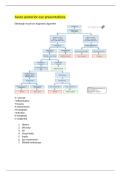
Edinburgh visual loss diagnostic algorithm
V- vascular
I-inflammation
T-trauma
A-autoimmune
M-metabolic
I-infection
N-neoplastic
C-congenital
1. History
2. Slit lamp
3. VA
4. Visual fields
5. Pupils
6. Eye movements
7. Dilated fundoscopy
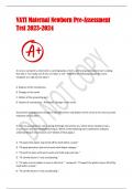
, CRAO BRAO
Symptoms Sudden painless Sudden painless
monocular visual loss monocular drop in vision
but vision often
unaffected
Signs -RAPD (profound) -RAPD often
-emboli -emboli at bifunicaction
-white, oedematous points
retina -white, oedematous
-cherry red spot section of retina
-disc pallor -Altitudinal defect
-retinal vasculature
narrowing
-holomhurst plaque – as
CRA emerges from disc
looks yellow
Management Initiate ocular massage Guidance less clear
wile px lies supine Same as CRAO
Same day emergency Px at risk of TIA/stroke
referral.
*bifurcation point: where branches off from the main arteriole route
*ocular massage: want to dislodge emboli
*lie supine: allows blood flow to be increased.
Management by ophthalmology:
Therapy is aimed at decreasing IOP
Acetazolamide
Intra-arterial fibrinolytic therapy
Aspirin- discourage platelet aggregation
Lifestyle advice
Discourage any further emboli from dislodging from carotid artery
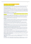
RVO:
Thrombus in CRV or a branch of- blocks venous system in the retina
If central- significant visual loss
If branch may not have any symptoms
Can have blurred vision, metamorphopsia and visual loss
CMO
CWS and oedema
Retinal whitening
Disc oedema
RAPD only in ischaemic CRVO
Ischaemic Non-ischaemic
RAPD No RAPD
Severe vision loss <6/60 Moderate visual loss >6/60
Lots of haemorrhages Fewer haemorrhages
Edinburgh visual loss diagnostic algorithm
V- vascular
I-inflammation
T-trauma
A-autoimmune
M-metabolic
I-infection
N-neoplastic
C-congenital
1. History
2. Slit lamp
3. VA
4. Visual fields
5. Pupils
6. Eye movements
7. Dilated fundoscopy
, CRAO BRAO
Symptoms Sudden painless Sudden painless
monocular visual loss monocular drop in vision
but vision often
unaffected
Signs -RAPD (profound) -RAPD often
-emboli -emboli at bifunicaction
-white, oedematous points
retina -white, oedematous
-cherry red spot section of retina
-disc pallor -Altitudinal defect
-retinal vasculature
narrowing
-holomhurst plaque – as
CRA emerges from disc
looks yellow
Management Initiate ocular massage Guidance less clear
wile px lies supine Same as CRAO
Same day emergency Px at risk of TIA/stroke
referral.
*bifurcation point: where branches off from the main arteriole route
*ocular massage: want to dislodge emboli
*lie supine: allows blood flow to be increased.
Management by ophthalmology:
Therapy is aimed at decreasing IOP
Acetazolamide
Intra-arterial fibrinolytic therapy
Aspirin- discourage platelet aggregation
Lifestyle advice
Discourage any further emboli from dislodging from carotid artery
RVO:
Thrombus in CRV or a branch of- blocks venous system in the retina
If central- significant visual loss
If branch may not have any symptoms
Can have blurred vision, metamorphopsia and visual loss
CMO
CWS and oedema
Retinal whitening
Disc oedema
RAPD only in ischaemic CRVO
Ischaemic Non-ischaemic
RAPD No RAPD
Severe vision loss <6/60 Moderate visual loss >6/60
Lots of haemorrhages Fewer haemorrhages