Pathologie 2
Table of Contents
Chapter 8, 9: Cardiovascular pathology ............................................................................. 1
Chapter 10: hematopoietic and lymphoid systems ............................................................. 9
Chapter 13: the lung ....................................................................................................... 11
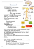
Chapter 21: brain and neurodegenerative diseases ......................................................... 15
Chapter 17: Gynaecopathology ....................................................................................... 20
Chapter 13: Pathology of the digestive tract ................................................................... 27
Chapter 21: Neuropathology........................................................................................... 32
Chapter 10: bone marrow ............................................................................................... 38
Chapter 8, 9: Cardiovascular pathology
Atherosclerosis
• Composition varies according to functional needs
• Certain disorders involve specific type of vessels
• Atherosclerosis: larger, muscular arteries
• Hypertension: small arterioles
Atherosclerosis: hardening of the arteries
- Vascular response to endothelial injury
- Causes:
o Hemodynamic disturbances
o Hypercholesterolemia
,Infrarenal aorta often involved with at → sheer stress bcs lost of blood flow and endothelial
damage
Reaction:
- Leukocytes to vessel wall
- Smooth muscle cells to stable effect
- Fatty streak
- Plaque
Danger → plaques obstruct lumen
Chronic inflammation is the corner stone
Component of plaques:
- 3 principal components of atherosclerotic plaques:
o Cells (SMC, macrophages, T-
cells)
o ECM (collagen, elastic
fibers, proteoglycans)
o Intracellular and
extracellular lipids
How often do they see a normal aorta →
never→ always fatty streaks
- Even children of 1 years old
You can get thrombosis from
atherosclerosis bcs blood can’t flow as well
Complications of Atherosclerosis
,Ischemic heart disease:
- Inbalance between demand of myocytes and supply of
oxygen
- Cardiac myocytes dependent on coronary blood flow for
survival
- Inadequate coronary perfusion relative to myocardial
demand
o Atherosclerotic occlusion of the coronary arteries
▪ Chronic
▪ Acute plaque changes → thrombosis
- Complications (3/4th of patients) such as myocardial
rupture
Pathophysiologic outcome depends on:
- Size affected vessel → in aorta not a big risk
- Size and stability of the plaque
- Degree plaque disrupts the vessel wall
Major clinical consequences of atherosclerosis = myocardial infarction, cerebral infarction,
aortic aneurysm, peripheral vascular disease
Plaques responsible for MI and ACS are often asymptomatic before the acute event.
Symptoms triggered due to thrombosis on a lesion that previously did not produce
significant luminal occlusion.
Thrombosis can also occur on an intact plaque.
Vulnerable plaques: large number of foam cells, thin fibrous cap, clusters of inflammatory
cells.
Inflammation destabilized the mechanical integrity of the plaque by increasing collagen
degradation. Statins reduce circulating cholesterol levels, but also reversal of endothelial
dysfunction.
Extrinsic factors also play a role → adrenergic stimulation → blood pressure increase, local
vasoconstriction (intense emotions) → increase mechanical stress → circadian periodicity of
heart attacks (6 am and 12 noon) → surge with walking and rising → blood pressure spikes +
heightened platelet reactivity.
Vulnerable plaques are at high risk of rupturing.
, Myocardial infarct:
- Zone of necrosis
- Zone that is spared because of blood
that comes from the lumen
- Exception of very narrow zone directly
subendocardially → direct blood flow from
ventricles
- First injury ischemic disease in
subendocardial zone → last to receive
blood flow and high pressure → impede
blood flow
- More prolongued ischemia → other
regions, driven by edema, ROS,
inflammatory mediators
- Infarct achieves its full extent into 3-6
hours
- Can have transmural infarction (full thickness, epicardial vessel
occlusion → atherosclerosis and acute plaque change with
trombosis
- Subendocardial infarction → spontaneous lyses of thrombus,
or therapy before it becomes transmural
Dominance of the coronary artery →
NBT stain:
- Normal: color purple
- Early disruption of
cardiomyotes → no stain
- First indication of myocardial
infarction: neutrophils
- After few days: granulation tissue
- Because of this they can see when the infarction occurred and if this was the reason.
For the death of the patient.
Complications of myocardial infarction:
Table of Contents
Chapter 8, 9: Cardiovascular pathology ............................................................................. 1
Chapter 10: hematopoietic and lymphoid systems ............................................................. 9
Chapter 13: the lung ....................................................................................................... 11
Chapter 21: brain and neurodegenerative diseases ......................................................... 15
Chapter 17: Gynaecopathology ....................................................................................... 20
Chapter 13: Pathology of the digestive tract ................................................................... 27
Chapter 21: Neuropathology........................................................................................... 32
Chapter 10: bone marrow ............................................................................................... 38
Chapter 8, 9: Cardiovascular pathology
Atherosclerosis
• Composition varies according to functional needs
• Certain disorders involve specific type of vessels
• Atherosclerosis: larger, muscular arteries
• Hypertension: small arterioles
Atherosclerosis: hardening of the arteries
- Vascular response to endothelial injury
- Causes:
o Hemodynamic disturbances
o Hypercholesterolemia
,Infrarenal aorta often involved with at → sheer stress bcs lost of blood flow and endothelial
damage
Reaction:
- Leukocytes to vessel wall
- Smooth muscle cells to stable effect
- Fatty streak
- Plaque
Danger → plaques obstruct lumen
Chronic inflammation is the corner stone
Component of plaques:
- 3 principal components of atherosclerotic plaques:
o Cells (SMC, macrophages, T-
cells)
o ECM (collagen, elastic
fibers, proteoglycans)
o Intracellular and
extracellular lipids
How often do they see a normal aorta →
never→ always fatty streaks
- Even children of 1 years old
You can get thrombosis from
atherosclerosis bcs blood can’t flow as well
Complications of Atherosclerosis
,Ischemic heart disease:
- Inbalance between demand of myocytes and supply of
oxygen
- Cardiac myocytes dependent on coronary blood flow for
survival
- Inadequate coronary perfusion relative to myocardial
demand
o Atherosclerotic occlusion of the coronary arteries
▪ Chronic
▪ Acute plaque changes → thrombosis
- Complications (3/4th of patients) such as myocardial
rupture
Pathophysiologic outcome depends on:
- Size affected vessel → in aorta not a big risk
- Size and stability of the plaque
- Degree plaque disrupts the vessel wall
Major clinical consequences of atherosclerosis = myocardial infarction, cerebral infarction,
aortic aneurysm, peripheral vascular disease
Plaques responsible for MI and ACS are often asymptomatic before the acute event.
Symptoms triggered due to thrombosis on a lesion that previously did not produce
significant luminal occlusion.
Thrombosis can also occur on an intact plaque.
Vulnerable plaques: large number of foam cells, thin fibrous cap, clusters of inflammatory
cells.
Inflammation destabilized the mechanical integrity of the plaque by increasing collagen
degradation. Statins reduce circulating cholesterol levels, but also reversal of endothelial
dysfunction.
Extrinsic factors also play a role → adrenergic stimulation → blood pressure increase, local
vasoconstriction (intense emotions) → increase mechanical stress → circadian periodicity of
heart attacks (6 am and 12 noon) → surge with walking and rising → blood pressure spikes +
heightened platelet reactivity.
Vulnerable plaques are at high risk of rupturing.
, Myocardial infarct:
- Zone of necrosis
- Zone that is spared because of blood
that comes from the lumen
- Exception of very narrow zone directly
subendocardially → direct blood flow from
ventricles
- First injury ischemic disease in
subendocardial zone → last to receive
blood flow and high pressure → impede
blood flow
- More prolongued ischemia → other
regions, driven by edema, ROS,
inflammatory mediators
- Infarct achieves its full extent into 3-6
hours
- Can have transmural infarction (full thickness, epicardial vessel
occlusion → atherosclerosis and acute plaque change with
trombosis
- Subendocardial infarction → spontaneous lyses of thrombus,
or therapy before it becomes transmural
Dominance of the coronary artery →
NBT stain:
- Normal: color purple
- Early disruption of
cardiomyotes → no stain
- First indication of myocardial
infarction: neutrophils
- After few days: granulation tissue
- Because of this they can see when the infarction occurred and if this was the reason.
For the death of the patient.
Complications of myocardial infarction: