APPP Cumulative Final Notes
CNS
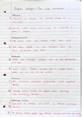
Learning Objectives:
Name the major bones of the skull/face, locate them on a drawing
Recognize principal parts of brain from axial, sagittal, coronal human brain image
Draw structure and function of dura
Name key blood vessels that supply head/brain, describe the importance of the circle of Willis
Describe formation and function of CSF
Identify the main anatomic components of brain (cortex, thalamus, cerebellum, brainstem,
hypothalamus)
Describe the function of parts of brain, spinal cord, brainstem, thalamus, hypothalamus, cortex
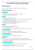
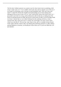
- Cranial bones: enclose, protect brain: frontal, temporal, occipital, parietal, sphenoid,
ethmoid
- Sutures connect bones: coronal, saggital, lamboid, squamosal
, - Foramen: nerves/blood vessels enter/exit
- Sinuses: paired cavities near nasal cavity: produce mucous, lighten skull, sound
transmission-should be filled w air
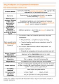
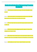
Cerebral Blood Flow
- To brain from carotid (L/R internal carotid arteries)
- Cerebral arterial circle of willis: allows 1 carotid to provide blood to whole brain
(aneurysms)
Strokes
- Ischemic: blockage
- Hemm: rupture→ brain bleed
- TIS: ischemic temp blockage
BBB
- Extends t/u CNS, protects from harmful substances (barrier to drug pen)
- Level of capp. (choroid plexus) endothelial cells w selective uptake→ tight junctions=
pass 2 membranes (lined w pumps)
- Astrocytes take up anything not permitted to pump back out
- Cranial meninges: cover brain: dura mater (fibrous), arachnoid, pia
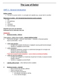
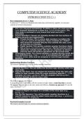
CSF
- Protective (shock absorber) and nutritive function→ allows structure
- Ventricles: cavities holding CSF → 2 lateral, one 3rd, one 4th (communicate w CSF in
cranial/spinal subarachnoid space)
- Formed via blood filtration in choroid plexus in ventricles
, - Cranial, spinal arachnoid villi= CSF absorption into jugular venous outflow
- Hydrocephalus: formation>clearance, increase intracranial P, loss of consc, decreased
brain fx
Brain
- Hemispheres linked by corpus callosum (mostly fibre)
- Gyri: convex fold, sulci: groove, fissure: deep sulci
- Gray matter: brain cells→ nuclei: brain cell groups
- White matter: fibres (myelinated axons)→ tracts: fiber collections (nerves going same
direction)
- Spinal cord: gray and white matter cts w medulla
➔ Segments (L/R spinal nerves arise): cervical, thoracic, lumbar, cauda equine (sacral
and coccygeal nerves don’t immediately leave vertebral canal
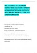
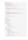
Spinal Cord Gray Matter
- Pos dorsal horn: receives/transmits sensory from dorsal roots (body input) to brain
(+initial integration) → dorsal root ganglia contain cell bodies (soma) of sensory nerve
- Anterior ventral horn: motor neuron cell bodies: signals to skel muscles via ventral roots
- Lateral horn: preganglionic symp motor neurons (ANS) (T1-L2)
, Ascending tracts: sensory
- Spinoothalamic: ant convey touch, pressure sensation, lat convey pain, temp→
thalamus
- Fasciculus gracilis and cuneatus: touch, proprioception, vibration, object quality
(→medulla)
- Spinocerebral tract: subconscious proprioception, motor control
Descending tracts: motor
- Corticospiral: motor impulses cortex→ motor neurons for consc mvmt
- Rubrospinal: motor impuses→ tone and posture
- Tectospinal: motor impulses tectum (MIDBRAIN)→control head movement in response
to stimuli
- Vestibulospinal: medulla→regulate balance
Spinal Cord
- Periphery sensory info→ brain
- Brain motor impulses→ periphery
- Integrates sensory, motor function via reflexes
Brain Stem- Medulla Oblongota
- Cts w spinal cord, allows communication
CNS
Learning Objectives:
Name the major bones of the skull/face, locate them on a drawing
Recognize principal parts of brain from axial, sagittal, coronal human brain image
Draw structure and function of dura
Name key blood vessels that supply head/brain, describe the importance of the circle of Willis
Describe formation and function of CSF
Identify the main anatomic components of brain (cortex, thalamus, cerebellum, brainstem,
hypothalamus)
Describe the function of parts of brain, spinal cord, brainstem, thalamus, hypothalamus, cortex
- Cranial bones: enclose, protect brain: frontal, temporal, occipital, parietal, sphenoid,
ethmoid
- Sutures connect bones: coronal, saggital, lamboid, squamosal
, - Foramen: nerves/blood vessels enter/exit
- Sinuses: paired cavities near nasal cavity: produce mucous, lighten skull, sound
transmission-should be filled w air
Cerebral Blood Flow
- To brain from carotid (L/R internal carotid arteries)
- Cerebral arterial circle of willis: allows 1 carotid to provide blood to whole brain
(aneurysms)
Strokes
- Ischemic: blockage
- Hemm: rupture→ brain bleed
- TIS: ischemic temp blockage
BBB
- Extends t/u CNS, protects from harmful substances (barrier to drug pen)
- Level of capp. (choroid plexus) endothelial cells w selective uptake→ tight junctions=
pass 2 membranes (lined w pumps)
- Astrocytes take up anything not permitted to pump back out
- Cranial meninges: cover brain: dura mater (fibrous), arachnoid, pia
CSF
- Protective (shock absorber) and nutritive function→ allows structure
- Ventricles: cavities holding CSF → 2 lateral, one 3rd, one 4th (communicate w CSF in
cranial/spinal subarachnoid space)
- Formed via blood filtration in choroid plexus in ventricles
, - Cranial, spinal arachnoid villi= CSF absorption into jugular venous outflow
- Hydrocephalus: formation>clearance, increase intracranial P, loss of consc, decreased
brain fx
Brain
- Hemispheres linked by corpus callosum (mostly fibre)
- Gyri: convex fold, sulci: groove, fissure: deep sulci
- Gray matter: brain cells→ nuclei: brain cell groups
- White matter: fibres (myelinated axons)→ tracts: fiber collections (nerves going same
direction)
- Spinal cord: gray and white matter cts w medulla
➔ Segments (L/R spinal nerves arise): cervical, thoracic, lumbar, cauda equine (sacral
and coccygeal nerves don’t immediately leave vertebral canal
Spinal Cord Gray Matter
- Pos dorsal horn: receives/transmits sensory from dorsal roots (body input) to brain
(+initial integration) → dorsal root ganglia contain cell bodies (soma) of sensory nerve
- Anterior ventral horn: motor neuron cell bodies: signals to skel muscles via ventral roots
- Lateral horn: preganglionic symp motor neurons (ANS) (T1-L2)
, Ascending tracts: sensory
- Spinoothalamic: ant convey touch, pressure sensation, lat convey pain, temp→
thalamus
- Fasciculus gracilis and cuneatus: touch, proprioception, vibration, object quality
(→medulla)
- Spinocerebral tract: subconscious proprioception, motor control
Descending tracts: motor
- Corticospiral: motor impulses cortex→ motor neurons for consc mvmt
- Rubrospinal: motor impuses→ tone and posture
- Tectospinal: motor impulses tectum (MIDBRAIN)→control head movement in response
to stimuli
- Vestibulospinal: medulla→regulate balance
Spinal Cord
- Periphery sensory info→ brain
- Brain motor impulses→ periphery
- Integrates sensory, motor function via reflexes
Brain Stem- Medulla Oblongota
- Cts w spinal cord, allows communication