Thorax II: Pleural/Pericardaic Cavities; Heart and Lungs
Key Points:
Surface markings of pleurae and lungs
Lung sounds
Surface markings and features of the heart
Heart sounds
Blood supply to the heart
Station 1: Pleurae and Lungs
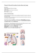
Pleural membranes are the fine serous membranes that line the thoracic cavity and
envelop the lungs.
The pleural cavity is a potential space between the parietal (lines thoracic wall) and
visceral layers (adheres to lung).
The pleural cavity is kept moist by a small volume of pleural fluid, each pleural
cavity is quite separate and contains no structures. The parietal pleura separates
the pleural cavity from the mediastinum
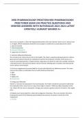
Parietal pleura – The parietal pleura is all one “sheet” but different parts are named
depending on where in the thoracic cavity they are lining.
Costal – Lines inner surface of ribs and intercostals
Diaphragmatic – Lines top of diaphragm
Mediastinal – Lines inner sides, either side of the heart
Cervical – Rises up to the neck, over apex of lung
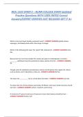
,The visceral pleura is firmly attached to the lungs, it is continuous with the
mediastinal pleura at the root of the lung.
Root of the lung—
Note the pulmonary ligament at the root of the lung (it is not a real ligament, rather
it is a fold of pleural membrane) that attaches the lung medially to the mediastinum.
Note that the pulmonary ligament lies inferior to the root of the lung
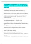
, Pleural Recesses –
During deep inspiration, the lungs fill the pleural cavities, but during quiet
respiration the lungs do not occupy some parts of the cavities. The spaces (seen
during quiet respiration) are called pleural recesses. There are two –
Costo-diaphragmatic recess
Costo-mediastinal recess –
Costo-diaphragmatic recess
Key Points:
Surface markings of pleurae and lungs
Lung sounds
Surface markings and features of the heart
Heart sounds
Blood supply to the heart
Station 1: Pleurae and Lungs
Pleural membranes are the fine serous membranes that line the thoracic cavity and
envelop the lungs.
The pleural cavity is a potential space between the parietal (lines thoracic wall) and
visceral layers (adheres to lung).
The pleural cavity is kept moist by a small volume of pleural fluid, each pleural
cavity is quite separate and contains no structures. The parietal pleura separates
the pleural cavity from the mediastinum
Parietal pleura – The parietal pleura is all one “sheet” but different parts are named
depending on where in the thoracic cavity they are lining.
Costal – Lines inner surface of ribs and intercostals
Diaphragmatic – Lines top of diaphragm
Mediastinal – Lines inner sides, either side of the heart
Cervical – Rises up to the neck, over apex of lung
,The visceral pleura is firmly attached to the lungs, it is continuous with the
mediastinal pleura at the root of the lung.
Root of the lung—
Note the pulmonary ligament at the root of the lung (it is not a real ligament, rather
it is a fold of pleural membrane) that attaches the lung medially to the mediastinum.
Note that the pulmonary ligament lies inferior to the root of the lung
, Pleural Recesses –
During deep inspiration, the lungs fill the pleural cavities, but during quiet
respiration the lungs do not occupy some parts of the cavities. The spaces (seen
during quiet respiration) are called pleural recesses. There are two –
Costo-diaphragmatic recess
Costo-mediastinal recess –
Costo-diaphragmatic recess