116A WEEK 11 – Introduction to cell death mechanisms
Learning outcomes:
1. Define the main mechanisms of cell death
2. Apoptosis: definition and relevance in physiology and diseases
3. Apoptosis: morphological features, the signalling pathway
4. Apoptosis: mechanisms of regulation
5. Type II: autophagic cell death
6. Type III: necrotic cell death
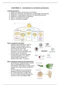
Type 1: Apoptotic cell death
- Happens automatically in cells at
the end of their lifespan: when
needed, apoptosis genes turn on
and direct the cell to die
- This is vital for life – without
apoptosis, old and damaged cells
would remain in the body
- An average of 50-70bn cells/day
undergo apoptosis in an adult
human body (average full
replacement every ~8 years)
Why is apoptosis important physiologically?
- Development: Apoptosis play a major
role in morphogenesis & tissue
remodelling: elimination of organs and
tissues only useful during embryonic
stages
- Ex: the tadpole tail is removed by
apoptosis during metamorphosis
- Ex: Individualization of digits that
occurs in many animals through the
removal of the inter-digital webs
, - Elimination of unnecessary cells
Why is apoptosis important?
- Defence mechanism
- Drugs used for cancer chemotherapy
- Hormones
Apoptosis in disease
- Associated with a number of pathological diseases
- Excessive apoptosis has been linked to neurodegenerative diseases
and organ failure after infarction or toxic insult
- Defective apoptosis seen in proliferative diseases i.e. cancer
Apoptosis induction – what causes it to begin?
- DNA damage to bases etc.
- Hypoxia (loss of oxygen)
- Oxidative stress (from free radicals)
- Death receptor ligands (usually on immune cell surface)
- Drug treatments (can refer to i.e.
chemotherapy or alcohol etc.)
3- Morphology and signalling in apoptosis
Hallmarks of apoptosis
- Cellular and nuclear shrinkage
- Disassembly into apoptotic bodies
- Chromatin condensation & DNA
fragmentation
- Mitochondrial membrane
permeabilization
- Cytochrome-c release from
mitochondria
- Caspases cascade activation
Cell plasma membrane is altered
- Externalisation of
phosphatidylserine
- Signal for phagocytosis
- Prevents immune response
Intracellular calcium in apoptosis
- Influx can trigger apoptosis
- Released from ER and taken up
by mitochondria
- Can be caused by improper
protein folding – trigger
- Expression of BcI2 trigger
calcium release and alters
membrane permeability
Learning outcomes:
1. Define the main mechanisms of cell death
2. Apoptosis: definition and relevance in physiology and diseases
3. Apoptosis: morphological features, the signalling pathway
4. Apoptosis: mechanisms of regulation
5. Type II: autophagic cell death
6. Type III: necrotic cell death
Type 1: Apoptotic cell death
- Happens automatically in cells at
the end of their lifespan: when
needed, apoptosis genes turn on
and direct the cell to die
- This is vital for life – without
apoptosis, old and damaged cells
would remain in the body
- An average of 50-70bn cells/day
undergo apoptosis in an adult
human body (average full
replacement every ~8 years)
Why is apoptosis important physiologically?
- Development: Apoptosis play a major
role in morphogenesis & tissue
remodelling: elimination of organs and
tissues only useful during embryonic
stages
- Ex: the tadpole tail is removed by
apoptosis during metamorphosis
- Ex: Individualization of digits that
occurs in many animals through the
removal of the inter-digital webs
, - Elimination of unnecessary cells
Why is apoptosis important?
- Defence mechanism
- Drugs used for cancer chemotherapy
- Hormones
Apoptosis in disease
- Associated with a number of pathological diseases
- Excessive apoptosis has been linked to neurodegenerative diseases
and organ failure after infarction or toxic insult
- Defective apoptosis seen in proliferative diseases i.e. cancer
Apoptosis induction – what causes it to begin?
- DNA damage to bases etc.
- Hypoxia (loss of oxygen)
- Oxidative stress (from free radicals)
- Death receptor ligands (usually on immune cell surface)
- Drug treatments (can refer to i.e.
chemotherapy or alcohol etc.)
3- Morphology and signalling in apoptosis
Hallmarks of apoptosis
- Cellular and nuclear shrinkage
- Disassembly into apoptotic bodies
- Chromatin condensation & DNA
fragmentation
- Mitochondrial membrane
permeabilization
- Cytochrome-c release from
mitochondria
- Caspases cascade activation
Cell plasma membrane is altered
- Externalisation of
phosphatidylserine
- Signal for phagocytosis
- Prevents immune response
Intracellular calcium in apoptosis
- Influx can trigger apoptosis
- Released from ER and taken up
by mitochondria
- Can be caused by improper
protein folding – trigger
- Expression of BcI2 trigger
calcium release and alters
membrane permeability