🔍 Chest Auscultation: A Structured Guide
for Medical Students
🩺 Introduction
Chest auscultation is a fundamental component of the respiratory examination.
Recognising various lung sounds is essential for identifying underlying pathologies and
guiding further diagnostic and management decisions.
This guide outlines a systematic approach to auscultation and describes the clinical features
and common causes of key respiratory sounds.
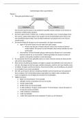
📍 Auscultation Technique
Systematic Approach
Auscultate in a side-to-side comparison, moving sequentially down the chest.
Listen to anterior, posterior, and lateral lung zones.
Use the diaphragm of the stethoscope.
Ask the patient to take deep breaths through the mouth.
Key Auscultation Sites
Anterior chest: Above and below the clavicles, mid-zone, and bases.
Posterior chest: Above the scapulae, interscapular region, mid-zone, and bases.
Lateral chest: In the mid-axillary line.
✅ Normal Breath Sounds
1. Vesicular Breathing
Low-pitched, soft sounds.
Heard primarily on inspiration.
Normal over most lung fields.
2. Bronchial Breathing
Louder, harsher sound (like over the trachea).
Equal inspiration and expiration with a pause in between.
Normal only near the trachea.
Abnormal elsewhere – suggests lung consolidation (e.g. pneumonia).
for Medical Students
🩺 Introduction
Chest auscultation is a fundamental component of the respiratory examination.
Recognising various lung sounds is essential for identifying underlying pathologies and
guiding further diagnostic and management decisions.
This guide outlines a systematic approach to auscultation and describes the clinical features
and common causes of key respiratory sounds.
📍 Auscultation Technique
Systematic Approach
Auscultate in a side-to-side comparison, moving sequentially down the chest.
Listen to anterior, posterior, and lateral lung zones.
Use the diaphragm of the stethoscope.
Ask the patient to take deep breaths through the mouth.
Key Auscultation Sites
Anterior chest: Above and below the clavicles, mid-zone, and bases.
Posterior chest: Above the scapulae, interscapular region, mid-zone, and bases.
Lateral chest: In the mid-axillary line.
✅ Normal Breath Sounds
1. Vesicular Breathing
Low-pitched, soft sounds.
Heard primarily on inspiration.
Normal over most lung fields.
2. Bronchial Breathing
Louder, harsher sound (like over the trachea).
Equal inspiration and expiration with a pause in between.
Normal only near the trachea.
Abnormal elsewhere – suggests lung consolidation (e.g. pneumonia).