Interpretation of the ECG
- The ECG is a fundamental part of cardiac assessment.
- Essential for investigating cardiac arrhythmias Ischaemia heart failure and other cardiac
disorders
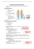
- The 12 lead ECG is made up of:
o Standard limb leads (I, II and III),
o ‘Augmented’ limb leads (aVR, aVL and
aVF)
o Six precordial leads (V1, V2, V3, V4, V5 and
V6).
-
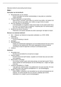
Conducting
- Starts at SA node
- Controlled by parasympathetic and sympathetic
nerves
- Send signals to the myocytes of the atria to
depolarise all the myocytes together
- this causes the atria to contract
- Pathway stops at AV node
- Travels to left and right bundle branches of the
purkinje fibres
- Depolarises the left and right ventricles which
then causes depolarisation
- The ECG is a fundamental part of cardiac assessment.
- Essential for investigating cardiac arrhythmias Ischaemia heart failure and other cardiac
disorders
- The 12 lead ECG is made up of:
o Standard limb leads (I, II and III),
o ‘Augmented’ limb leads (aVR, aVL and
aVF)
o Six precordial leads (V1, V2, V3, V4, V5 and
V6).
-
Conducting
- Starts at SA node
- Controlled by parasympathetic and sympathetic
nerves
- Send signals to the myocytes of the atria to
depolarise all the myocytes together
- this causes the atria to contract
- Pathway stops at AV node
- Travels to left and right bundle branches of the
purkinje fibres
- Depolarises the left and right ventricles which
then causes depolarisation