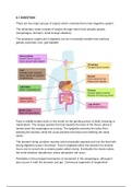
6.1 DIGESTION
There are two major groups of organs which comprise the human digestive system:
The alimentary canal consists of organs through which food actually passes
(oesophagus, stomach, small & large intestine)
The accessory organs aid in digestion but do not actually transfer food (salivary
glands, pancreas, liver, gall bladder)
Food is initially broken down in the mouth by the grinding action of teeth (chewing or
mastication). The tongue pushes the food towards the back of the throat, where it
travels down the esophagus as a bolus. The epiglottis prevents the bolus from
entering the trachea, while the uvula prevents the bolus from entering the nasal
cavity.
The stomach lining contains muscles which physically squeeze and mix the food with
strong digestive juices ('churning’). Food is digested within the stomach for several
hours and is turned into a creamy paste called chyme. Eventually the chyme enters
the small intestine (duodenum) where absorption will occur.
Peristalsis is the principal mechanism of movement in the oesophagus, although it
also occurs in both the stomach and gut. Continuous segments of longitudinal
,smooth muscle rhythmically contract and relax. Food is moved unidirectionally along
the alimentary canal in a caudal direction (mouth to anus).
Segmentation involves the contraction and relaxation of non-adjacent segments of
circular smooth muscle in the intestines. Segmentation contractions move chyme in
both directions, allowing for a greater mixing of food with digestive juices .While
segmentation helps to physically digest food particles, its bidirectional propulsion of
chyme can slow overall movement.
The stomach contains gastric glands which release digestive acids to create a low
pH environment (pH ~2). The acidic environment functions to denature proteins and
other macromolecules, aiding in their overall digestion. The stomach epithelium
contains a mucous membrane which prevents the acids from damaging the gastric
lining. The pancreas releases alkaline compounds (e.g. bicarbonate ions), which
neutralise the acids as they enter the intestine.
The liver produces a fluid called bile which is stored and concentrated within the gall
bladder prior to release into the intestine. Bile contains bile salts which interact with
fat globules and divide them into smaller droplets (emulsification). The emulsification
of fats increases the total surface area available for enzyme activity (lipase).
Enzymes are biological catalysts which speed up the rate of a chemical reaction (i.e.
digestion) by lowering activation energy. Enzymes allow digestive processes to
therefore occur at body temperatures and at sufficient speeds for survival
requirements. Enzymes are specific for a substrate and so can allow digestion of
certain molecules to occur independently in distinct locations.
The small intestine is composed of four main tissue layers, which are (from outside
to centre):
★ Serosa – a protective outer covering composed of a layer of cells reinforced
by fibrous connective tissue
★ Muscle layer – outer layer of longitudinal muscle (peristalsis) and inner layer
of circular muscle (segmentation)
★ Submucosa – composed of connective tissue separating the muscle layer
from the innermost mucosa
★ Mucosa – a highly folded inner layer which absorbs material through its
surface epithelium from the intestinal lumen
, The epithelial lining of villi contains several structural features which optimise its
capacity to absorb digested materials. Occluding associations between the plasma
membrane of two adjacent cells, creating an impermeable barrier. They keep
digestive fluids separated from tissues and maintain a concentration gradient by
ensuring one-way movement.
Microvilli borders significantly increase surface area of the plasma membrane
(>100×), allowing for more absorption to occur. The membrane will be embedded
with immobilised digestive enzymes and channel proteins to assist in material
uptake.
Epithelial cells of the intestinal villi will possess large numbers of mitochondria to
provide ATP for active transport mechanisms. ATP may be required for primary
active transport (against gradient), secondary active transport (co-transport) or
pinocytosis.
Pinocytosis (‘cell-drinking’) is the non-specific uptake of fluids and dissolved solutes
(a quick way to translocate in bulk). These materials will be ingested via the breaking
and reforming of the membrane and hence contained within a vesicle.
A transport protein couples the active translocation of one molecule to the passive
movement of another (co-transport). Glucose and amino acids are co-transported
across the epithelial membrane by the active translocation of sodium ions (Na+).
Channel proteins help hydrophilic food molecules pass through the hydrophobic
portion of the plasma membrane. Certain monosaccharides (e.g. fructose), vitamins
and some minerals are transported by facilitated diffusion.
Water molecules will diffuse across the membrane in response to the movement of
ions and hydrophilic monomers (solutes).
Hydrophobic materials (e.g. lipids) may freely pass through the hydrophobic portion
of the plasma membrane. Once absorbed, lipids will often pass first into the lacteals
rather than being transported via the blood.
Endocytosis involves the invagination of the plasma membrane to create an internal
vesicle containing extracellular material. Vesicle formation requires the breaking and
reforming of the phospholipid bilayer and hence is an energy-dependent process. In
the intestines, vesicles commonly form around fluid containing dissolved materials
(pinocytosis – cell ‘drinking’). Pinocytosis allows materials to be ingested en masse
and hence takes less time than shuttling via membrane proteins.