4.2.1 – Principles of Organisation
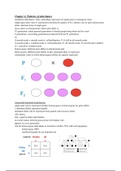
o A cell is the basic building block of all organisms.
o An organelle is a specialised unit within a cell which performs a specific function.
o A tissue is a group of cells working together to perform a shared function, and often with a similar structure.
o An organ is a structure made up of groups of different tissues, working together to perform specific functions.
o An organ system is a group of organs with related functions, working together to perform certain functions within the body.
4.2.2.1 – The Human Digestive System
o An ENZYME is a protein which acts as a biological catalyst.
• A substrate is what the enzyme breaks down.
• The product is what the substrate has broken down into.
o A CATALYST is a substance which speeds up the rate of a chemical reaction, but is not itself changed by the reaction.
• All enzymes are proteins, but not all proteins are enzymes.
• Catalase is the enzyme which breaks down hydrogen peroxide into water + oxygen.
o All enzymes contain an ACTIVE SITE where the reaction occurs which is complementary to the substrate - known as the
LOCK AND KEY THEORY.
o An enzyme’s optimum temperature is the temperature at which enzymes work best at.
• In the human body, an enzyme’s optimum temperature is 37 degrees Celsius, which is body temperature.
o The rate of reaction is the speed of the reaction per unit time.
o At low temperatures, enzymes are DEACTIVATED or they work slower.
o When enzymes are at really high temperatures, the active site changes permanently per minute, causing them to be rendered
unusable, which is called DENATURING.
• Retaining high temperatures can cause toxin production.
• Deactivated enzymes can work again when temperature rises, but denatured enzymes are essentially dead, and cells
have to make new ones.
o As temperature rises from the point of deactivation: o As temperature rises past the optimum temperature
• Greater kinetic energy towards the point of denaturing:
• Increased collisions between the enzymes and • Active site starts to change shape – begins
substrates denaturing
• Higher rate of reaction • Less successful collisions due to the less
complementary shape
• Lower rate of reaction
o Enzymes prefer to work at a specific pH, called the optimum pH – above or below the optimum pH, the enzyme will become
denatured.
• Enzymes in the stomach work at an optimum pH of 2, the same as the hydrochloric acid present.
o Carbohydrates, proteins, lipids (or fats) are major nutrients that we need in large quantities & are gained by eating them.
o They’re broken down first and then reassembled into our own carbohydrates, proteins and lipids. This is because:
• most of the molecules in food are too large to pass through the absorbing surface of the gut wall
• the carbohydrates, proteins and lipids are reassembled in the form required, rather than other animal or plant versions
o This follows the three steps of digestion:
• Digestion – breaking down of large insoluble molecules into smaller soluble molecules.
▪ This involves emulsification where the gall bladder releases a chemical called bile which neutralises stomach
acid and breaks down the large molecules specifically for the digestion of lipids.
• Absorption – transfer of smaller soluble molecules into the blood plasma.
• Assimilation – making new proteins by reassembling the smaller soluble molecules.
o There are two types of digestion: Physical (biting with the teeth, churning with the stomach muscles) & Chemical (use of acid
in the stomach).
o Carbohydrate nutrient:
• Function: source of energy → glucose is the main respiratory substrate.
• Found in: starch (potatoes, rice and wheat products, bread, cereals and pasta) / sugars (fruit, smoothies, fizzy drinks,
chocolate and sweets).
o Proteins:
• Function: growth and repair.
• Found in: meat, eggs, cheese, beans, nuts and seeds.
o Lipids:
• Function: energy, makes up part of the cell membrane so it is essential for normal growth.
• Found in: butter and margarine, meat and processed meat, plant oils, oily fish, nuts and seeds.
o The mouth releases the enzyme AMYLASE which breaks down the substrate STARCH into the product MALTOSE.
o The stomach releases the enzyme PEPSIN which breaks down the substrate PROTEIN into the product AMINO ACIDS.
o The small intestine (/pancreas) releases:
• The enzyme LIPASE which breaks down the substrate LIPIDS into the product FATTY ACIDS + GLYCEROL.
• The enzyme AMYLASE which breaks down the substrate STARCH into the product MALTOSE.
• The enzyme PROTEASE which breaks down the substrate PROTEIN into the product AMINO ACIDS.
• The enzyme MALTASE which breaks down the substrate MALTOSE into the product GLUCOSE.
o The large intestine carries out the absorption of water.
o Testing for starch:
, • If starch is present, it changes IODINE solution from orangey-brown to blue black.
o Testing for glucose:
• Dissolve the solution in a test tube of water.
• Add a few drops of BENEDICT solution and then HEAT.
• If glucose is present in the solution, it will change from blue to brick red. The higher the colour, from blue to green to
orange to red, the more glucose that is present.
o Testing for lipids:
• Dissolve in water and add a few drops of ALCOHOL. If lipids are present, it will form a cloudy white emulsion.
o Test for proteins:
• Add the protein sample and dissolve in water. Add a few drops of BIURET solution and SHAKE.
• If the colour changes from blue to violet then proteins are present.
4.2.2.2 – The Heart and Blood Vessels + 4.2.2.3 – The Blood
o Blood is made up of: red blood cells, white blood cells, plasma and platelets.
o Red blood cells:
• Have a biconcave shape and no nucleus.
• Carries oxygen – contains haemoglobin to do this.
• Oxygen + Haemoglobin → Oxyhaemoglobin
• Oxygen is released from the oxyhaemoglobin into the cells and the haemoglobin reverts back to normal until it picks up
oxygen again.
o White blood cells:
• The largest blood cell – large nucleus and can change shape.
• Protects the body from harmful pathogens and invading microbes.
• Phagocyte white blood cells: engulfs the pathogen using enzymes called lysozymes to break it down and destroy it.
• Lymphocyte white blood cells: produces antibodies to neutralise antigens produced by the pathogen / antitoxins to
neutralise toxins produced by the pathogen.
o Platelets:
• Cell fragments broken off from larger cells.
• Smallest blood cells and have no nucleus.
• Platelets help to pick up tiny fibres that form a net at the site of a cut.
• Red blood cells are trapped in this which forms a blood clot.
• The clot dries and forms a scab which protects the cut whilst new skin grows.
o Plasma is the liquid part of the blood which carries: RBCs, WBCs, platelets, amino acids, glucose, CARBON DIOXIDE and
urea.
o OXYGENATED BLOOD is blood which contains a higher concentration of oxygen than the concentration of carbon dioxide.
o DEOXYGENATED BLOOD is blood which contains a higher concentration of carbon dioxide than the concentration of
oxygen.
o Pulmonary is a term associated with the lungs.
o A VEIN usually carries blood towards the heart. o An ARTERY usually carries blood away from the
• Usually low in oxygen and a purple-red colour. heart.
• Thin walls and valves which open to let blood • Usually high in oxygen with a bright red colour.
through – no pulse unlike arteries. • Stretches as blood is forced through it and returns
• If blood flows backwards, the valves close to to its normal shape afterwards.
prevent the BACKFLOW of blood – blood is then • Thick walls containing muscle and elastic fibres.
squeezed back to the heart using the skeletal
muscles.
o CAPILLARIES form a huge network of tiny vessels which link arteries and veins.
• Narrow thin walls allow substances (oxygen, glucose etc) to diffuse easily out of the blood into the cells.
• Substances produced by cells like carbon dioxide can pass easily into the blood through capillary walls.
o Oxygen diffuses from the alveoli into the red blood cells. Carbon dioxide diffuses from the plasma into the alveoli.
o The circulatory system consists of two parts: the heart & the blood vessels (veins, capillaries and arteries) – the two parts
make it a DOUBLE-CIRCULATORY SYSTEM.
o The function of a VALVE is to prevent the back flow of blood.
o The PULMONARY VEIN carries oxygenated blood from the lungs to the heart.
o The AORTA carries oxygenated blood from the heart to the rest of the body.
o The VENA CAVA carries deoxygenated blood from the body to the heart.
o The PULMONARY ARTERY carries deoxygenated blood from the heart to the lungs.
1. Deoxygenated blood enters the vena cava.
2. The blood enters through the right atrium and forces its way into the tricuspid valve which then closes preventing
backflow, and the blood then exits into the right ventricle.
3. Enters the right semi-lunar valve and exits through the pulmonary artery where the heart contracts and sends the blood to
the lungs where it gets oxygenated.
4. Oxygenated blood travels through the lungs into the pulmonary vein, then forces its way into and exits the bicuspid valve
causing it to close preventing back flow, into the left ventricle.
5. It enters the left semi-lunar valve and the heart contracts, causing the oxygenated blood to exit the heart through the aorta
towards the body.
6. Oxygen diffuses into the body cells and the blood becomes deoxygenated, then picks up carbon dioxide into the plasma.
7. The deoxygenated blood travels from the body to the Vena Cava.
o The SEPTUM crosses through the middle of the heart and prevents deoxygenated and oxygenated blood from mixing.
o The heart is made up of cardiac muscle meaning it doesn’t need to take a break.