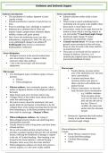
Entheses and Enthesis Organs
Entheses introduction Stress concentration
The attachment of a tendon, ligament or joint Entheses attaches tendon (soft) to bone
capsule to bone (hard)
Smooth and effective transfer of muscle force to When trying to attach something hard to
bone. something soft, the point in the middle where
Prone to pathology and a challenge to engineer! these attach is most likely to fail
Entheses are a common site of ‘Over use Locomotion, results in the angle between the
Injuries’ (sport- jumpers knee, throwers elbow, tendon or bone which is moving relative to
Achilles, rotator cuff, groin strains) one and another insertional angle change
Don’t know the underlying cause. (are they insertional angle change makes
inflammatory, degenerative, both? We dk), so it attachment site potentially weaker
is any pathological change to an enthesis Ground reaction forces Contact with the
Enthesopathy (also known as insertional ground causes movement of the soft tissue,
tendinopathies, enthesitis) these are also focussed at the tissue interface
(at attachment site).
These give an increased risk for rupture or
Stress dissipation damage of entheses. However, it is
Strongest structure in the muscle-tendon-bone anatomically adapted (stress dissipation) to
unit, more likely to have a rupture in these prevent the likelihood of injury.
structures rather than entheses.
^ due to the macroscopic and microscopic
adaptations
Macroscopic
Microscopic Tendon flaring- increase surface
Two histological types of entheses (types of tissue area of the attachment site- decrease
present) stress concentration
o Fibrous Common attachment sites-
increases surface area (seen at
o Fibrocartilaginous
elbow and foot with achilles and
plantar fascia)
Fibrous entheses- most commonly present, where Fibrous connections- to share the
tendon or ligament attaches to the diaphysis (shaft of load, decrease stress concentration
the bone) - e.g. Achilles continues
Quite broad, span out to increase surface area from calcaneus all the
Collagen fibres of the tendon insert almost directly way over the back of the
under the underlying bone heel and into the sole of
Not much is know about the attachment, but most the foot. Fibres of
people think the mechanism of attachment is by the Achilles also insert and
collagen fibres of the tendon inserting directly into the anchor into the calcaneal
bone underlying it, so the fibres penetrate into the fat pad
bone itself for anchorage – Sharpey’s fibres
Retinacula- ligaments that hold tendons
Fibrocartilaginous entheses- has a plug of closely to the bone minimises
fibrocartilage between tendon and underlying bone insertional angle change
(intermediate material)
Usually found on ends of long bones and short bones
of hand and feet
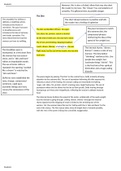
Not as soft as tendon and not as hard as bone, so Fibrocartilaginous Entheses
provides a gradual transition between the soft and hard 4 zones
tissues, reducing the likelihood of damage or rupture Dense fibrous connective tissue
at the site Gradual bending - fibre bending is not Uncalcified fibrocartilage
focused at the tissue interface= reduced fraying + Calcified fibrocartilage
dissipates stress away from the tissue interface Bone
Fibrocartilaginous entheses are much more prone to
overuse injuries, it is also more widely studied
Entheses introduction Stress concentration
The attachment of a tendon, ligament or joint Entheses attaches tendon (soft) to bone
capsule to bone (hard)
Smooth and effective transfer of muscle force to When trying to attach something hard to
bone. something soft, the point in the middle where
Prone to pathology and a challenge to engineer! these attach is most likely to fail
Entheses are a common site of ‘Over use Locomotion, results in the angle between the
Injuries’ (sport- jumpers knee, throwers elbow, tendon or bone which is moving relative to
Achilles, rotator cuff, groin strains) one and another insertional angle change
Don’t know the underlying cause. (are they insertional angle change makes
inflammatory, degenerative, both? We dk), so it attachment site potentially weaker
is any pathological change to an enthesis Ground reaction forces Contact with the
Enthesopathy (also known as insertional ground causes movement of the soft tissue,
tendinopathies, enthesitis) these are also focussed at the tissue interface
(at attachment site).
These give an increased risk for rupture or
Stress dissipation damage of entheses. However, it is
Strongest structure in the muscle-tendon-bone anatomically adapted (stress dissipation) to
unit, more likely to have a rupture in these prevent the likelihood of injury.
structures rather than entheses.
^ due to the macroscopic and microscopic
adaptations
Macroscopic
Microscopic Tendon flaring- increase surface
Two histological types of entheses (types of tissue area of the attachment site- decrease
present) stress concentration
o Fibrous Common attachment sites-
increases surface area (seen at
o Fibrocartilaginous
elbow and foot with achilles and
plantar fascia)
Fibrous entheses- most commonly present, where Fibrous connections- to share the
tendon or ligament attaches to the diaphysis (shaft of load, decrease stress concentration
the bone) - e.g. Achilles continues
Quite broad, span out to increase surface area from calcaneus all the
Collagen fibres of the tendon insert almost directly way over the back of the
under the underlying bone heel and into the sole of
Not much is know about the attachment, but most the foot. Fibres of
people think the mechanism of attachment is by the Achilles also insert and
collagen fibres of the tendon inserting directly into the anchor into the calcaneal
bone underlying it, so the fibres penetrate into the fat pad
bone itself for anchorage – Sharpey’s fibres
Retinacula- ligaments that hold tendons
Fibrocartilaginous entheses- has a plug of closely to the bone minimises
fibrocartilage between tendon and underlying bone insertional angle change
(intermediate material)
Usually found on ends of long bones and short bones
of hand and feet
Not as soft as tendon and not as hard as bone, so Fibrocartilaginous Entheses
provides a gradual transition between the soft and hard 4 zones
tissues, reducing the likelihood of damage or rupture Dense fibrous connective tissue
at the site Gradual bending - fibre bending is not Uncalcified fibrocartilage
focused at the tissue interface= reduced fraying + Calcified fibrocartilage
dissipates stress away from the tissue interface Bone
Fibrocartilaginous entheses are much more prone to
overuse injuries, it is also more widely studied