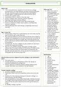
Osteoarthritis
What is OA?
“OA is characterised by a progressive loss of articular cartilage Why study OA?
accompanied by new bone formation and often synovial proliferation There are no cures or
that may culminate in pain, loss of joint function and disability” disease-modifying
Variable degrees of inflammation but without systemic effects drugs(DMOAD)to stop,
Primary and secondary forms slow, or reverse OA
Osteoarthritis (OA) is a disease of the entire joint progression
Involves cartilage, bone, synovium, meniscus, ligaments No way to build OA
Pathology leads to pain and joint stiffness The heterogeneous
Most commonly affected Joints: knees, hips, hands & spine nature of the disease,
Disease is initiated by multiple factors; genetics, developmental, lack of early diagnostic
traumatic, metabolic.... markers, mismatched
Differing etiologies but similar biologic, morphologic and clinical preclinical animal
outcomes models and clinical
populations, and the
complex role of many
targets of interest
Hip vs. Knee OA The OA pain experience
The prevalence of hip OA is similar between men and women, but that is very complex,
knee OA affects more women than men including both
The anatomical differences between the hip and knee joint are likely to peripheral and central
underpin why malalignment is risk factor for knee OA, but not hip OA pro-cesses and may
People with hip OA tend to be younger (60.4 years) than people with include nociceptive,
knee OA (66.3 years and have shorter duration of symptoms at the time inflammatory, and
of presentation neuropathic pain
Of individuals >55 years, multiple-site joint problems are much more Ove 50,000 knee and
common than single joint problems nearly 55,000 hip
Hip OA is less common among Chinese than US White women replacements in 2020
whereas knee OA is more common in Chinese than US White women Rare to have OA in just
Obesity, a strong risk factor for clinical knee OA progression, has one joint
generally not been found to be a strong risk factor for symptomatic or
radiographic hip OA disease progression
Epidemiology
Age
Does the disease process originate from the cartilage or the subchondral Obesity
bone? Overuse
Not really known Trauma: PTOA
OA Does the increase in SCB thickness reflects the changes in Deformity
mechanical loading due to cartilage loss? Instability
Or Does reduction in SCB bone stiffness during loading increase Genetic predisposition
cartilage deformation and contribute to OA development? Infection
Crystal deposition
OA has high
Normal articular cartilage heritability, estimated
avascular, alymphatic and aneural between 40%and 60%
chondrocyte which rises from chondroblast which comes from stem A paper said OA is a
cell disease of the whole
chondrocyte is the permenant phenotype of articular cartilage, it is person, due to the co-
characterised by type IIB collagen morbidities associated
these chondroblasts can also produce hypertrophic chondrocytes (type with it: depression,
10 collagen) found in growth plates, but this is hypertrophic cartilage, anxiety, pain
which will then die and be replaced by bone when growth plates are processing, pain
closed sensitisation
So, Healthy cartilage= type IIB
, Normal articular cartilage
Articular cartilage in OA
Metabolism is a highly
regulated balance
between synthesis and
degradation of the
various matrix
components
Very slow rate of getting
rid of old and making
new
OA: Biosynthetic
anabolic activity is
WEIGHT: unable to keep pace with
Water - 70% the degradative catabolic
Collagens - 20% activity and degeneration
Proteoglycans – 7% of the tissue results
Cells – 2% Thus, your body is meant
Other Proteins – 1% to keep a balance
(homeostasis of
Type II collagen (94%): degradation and
major fibrillar collagen that provides tensile strength. Form an biosynthesis, but in OA
organised fibrillar meshwork degradative catabolic
activity outweighs
Chondrocytes biosynthetic anabolic
react to mechanical stimuli and exist in a low oxygen tension activity
environment (HIF1α) One theory is that
aberrant distribution of
Aggrecan: forces in cartilage leads
A very strong affinity for binding water. Responsible for the to altered
compressive stiffness of AC mechanotransduction in
the chondrocytes and
Maturation and growth subsequent:
When born, articular cartilage was unorganised, then with maturation it o Activation of
becomes very organised, with different zones. Chondrocytes are flat at catabolic and
superficial zone then become rounder and more columnar as you go to inflammatory
the deep zone. genes
Once maturity is reached, chondrocytes do not divide again unless o Deregulated
pathology occurs cartilage matrix
synthesis
o Structural and
functional
changes to the
subchondral
bone
OA is not simply the
consequence of aging
o Some elderly
have perfect
cartilage, but
aging is a risk
factor