lOMoAR cPSD| 54901389
Introduction To The Human Body
Readings – Chapter 1
Anatomy – the study of internal and external body structures and their physical
relationships among other body parts.
Physiology – the study of how living organisms perform their functions.
Medical terminology – involves using word roots, prefixes, suffixed and combining
forms to build terms related to the body in health and disease.
Eponyms – anatomical structures and clinical conditions names after the discoverer
or most famous victim.
International Anatomical Terminology (Terminologia Anatomica or TA) – The
Federative Committee on Anatomical Terminology and 56 member associations on
the International Associations of Anatomists publishes this as a worldwide official
standard of anatomical vocabulary in 1998.
Principle of complementarity of structure and function – all specific functions are
performed by specific structures and the form of a structure related to its function.
Anatomy
Gross Anatomy (macroscopic) involves examining relatively large structures.
• Surface anatomy – study of general form and superficial markings.
• Regional anatomy – focuses on anatomical organisation of specific areas of
the body.
• Systemic anatomy – the study of the structure of organ systems which are
groups of organs that function together in a coordinated manner.
• Clinical anatomy – includes a number of specialities important in clinical
practice such as pathological anatomy, radiographic anatomy and surgical
anatomy.
• Developmental anatomy – describes the changes in form that take place
between conception and adulthood.
Microscopic Anatomy deals with structures we cannot see without magnification.
Cytology – the study of the internal structure of individual cells.
• Histology – the examination of tissues (groups of specialised cells and cell
products that work together to perform specific functions).
• Organs – tissues that combined to form organs (not always microscopic
anatomy).
Physiology
• Cell physiology – the study of the functions of cells, looking at events
involving the molecules and atoms important to life.
• Organ physiology – the study of the function of specific organs (i.e. cardiac
physiology)
• Systemic physiology – includes all aspects of the functioning of specific organ
systems.
Downloaded by Beavan Chomba ()
, lOMoAR cPSD| 54901389
• Pathological physiology – the study of the effects of diseases on organ
functions or system functions.
Signs – an objective disease indication such as fever.
Symptoms – a subjective disease indication such as tiredness.
Scientific method - a system of advancing knowledge that begins by proposing a
hypothesis to answer a question, and then testing that hypothesis with data
collected.
Levels of organisation progress from molecules to a complete organism
The Chemical Level – Atoms
Smallest stable units of matter.
• Combine to form molecules.
The Cellular Level – Cells
• Smallest living units in the body.
• Complex molecules cab form types of larger structures called organelles.
• Each organelle has a specific function in a cell.
• Cells perform 8 life functions: maintaining boundaries, movement,
responsiveness, digestion, metabolism, excretion, reproduction and growth.
The Tissue Level – Tissue
• A group of cells working together to perform one or more specific functions.
• Four main types of tissue: epithelial tissue, connective tissue, muscle tissue
and nervous tissue.
The Organ Level – Organs
• Made of two or more tissues working together to perform specific functions.
The Organ System Level – Organ system
• A group of organs interacting to perform a particular function forms and
organ system.
The Organism Level – Organism
• The highest level of organisation that we consider.
There is a written example on p.33 and a visual representation on p.35.
Homeostasis – refers to the existence of a stable internal environment.
Homeostatic regulation – is the adjustment of physiological systems to preserve
homeostasis. Contains two parts:
1. Autoregulation – a process that occurs when a tissue, organ or organ system
adjusts in response to some environmental change.
2. Extrinsic regulation – is a process that results from activities of the nervous
system or endocrine system. These organ system detect environmental
change and send an electric signal (nervous) or chemical messenger
(endocrine) to control or adjust the activities of one or more systems
simultaneously.
Homeostatic regulatory mechanism contains three parts:
1. Receptor – a sensor that is sensitive to a stimulus or particular environmental
change.
, lOMoAR cPSD| 54901389
2. Controls centre – receives and processes the information supplied by the
receptor and sends out commands.
3. Effector - a cell or organ that responds to the commands of the control centre
and whose activity either opposes or enhances the stimulus.
Negative feedback
• Primary mechanism of homeostatic regulation as it is a way of counteracting
change.
• Ignores minor variation as it has a normal range rather than fixed value.
Positive feedback
• Produces a response that exaggerates or enhances the original change in
conditions.
• Positive feedback loop – when a potentially dangerous process needs to be
completed quickly to restore homeostasis. E.g. blood clotting.
State of equilibrium – when opposing process or forces are in balance. E.g. rate of
heat loss equals rate of heat production.
Dynamic equilibrium – keeps vital conditions within a normal range of values.
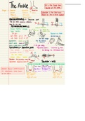
Body regions, anatomical positions and directions and body sections
Superficial anatomy
• Involves locating structures on or near body surface.
Anatomical landmarks
• Anatomical position – feet together, hands by side with palms facing forward.
• Seen from front – anterior view.
• Seen from the back – posterior view.
• Lying down in anatomical position face up – supine.
• Lying down in anatomical position face down – prone.
Anatomical regions
• Used when describing broader regions of the body.
• Abdominopelvic quadrants – formed by a pair of imaginary line that intersect
at umbilicus (navel).
• 9 Abdominopelvic regions – more precise terms to describe location and
orientation of internal organs.
• Axial part – head, neck and trunk.
• Appendicular part – consisting of the limbs.
• Anatomical relationships – relationships between the Abdominopelvic
quadrants and regions and the location of internal organs.
There is a written example on p.42 and a visual representation on p.42.
Anatomical directions
• Lateral – away from the midline.
• Medial – toward the midline.
• Proximal – toward the point of attachment of a limb to the trunk.
• Distal – away from the point of attachment of a limb to the trunk.
Downloaded by Beavan Chomba ()
, lOMoAR cPSD| 54901389
• Cranial or cephalic – towards the head.
• Caudal – towards the tail.
• Posterior or dorsal – the back or back surface.
• Anterior or ventral – the front or front surface.
• Superficial – at, near or relatively close to the body surface.
• Deep – toward the interior of the body; father from the surface.
• Lateral view – from the side.
• Superior – above, at a higher level. Inferior, below, at a lower level.
Sectional anatomy
• Slice through a three dimensional object is a section.
• Can be described in reference to three sectional planes.
• A plane is an axis.
• Transverse or horizontal plane – right angles to the long axis. A cut in this
place is called a transverse or cross section (top and bottom).
• Frontal plane (front and back) and sagittal plane (left and right) are parallel to
the long axis of the body.
Body cavities of the trunk Three
major regions:
1. Thoracic.
2. Abdominal.
3. Pelvic.
Body cavities – are closed, fluid filled and lined by a thin tissue layer called a serous
membrane or serosa.
Two main functions:
1.Protect delicate organs from shocks and impacts.
2.Permit significant changes in the size and shape of internal organs.
Diaphragm – separates the abdominopelvic and thoracic cavities. Viscera –
the internal organs that are enclosed by these cavities.
Thoracic cavity
• Contain the lungs and heart; organs of the respiratory, cardiovascular and
lymphatic systems.
• Pleural cavities – left and right cavities, which are separated by a mass of
tissue called the mediastinum.
• Mediastinum surrounds and supports esophagus, trachea and thymus.
• The mediastinum contains he pericardial cavity, a small cavity that surrounds
the heart.
• Pericardium is the serous membrane associated with the heart.
• Each pleural cavity surrounds a lung, and is lined by a serous membrane
called pleura.
• Visceral pleura covers outer surface of lung and parietal pleura covers
mediastinal surface and inner body wall.
Abdominopelvic cavity
Introduction To The Human Body
Readings – Chapter 1
Anatomy – the study of internal and external body structures and their physical
relationships among other body parts.
Physiology – the study of how living organisms perform their functions.
Medical terminology – involves using word roots, prefixes, suffixed and combining
forms to build terms related to the body in health and disease.
Eponyms – anatomical structures and clinical conditions names after the discoverer
or most famous victim.
International Anatomical Terminology (Terminologia Anatomica or TA) – The
Federative Committee on Anatomical Terminology and 56 member associations on
the International Associations of Anatomists publishes this as a worldwide official
standard of anatomical vocabulary in 1998.
Principle of complementarity of structure and function – all specific functions are
performed by specific structures and the form of a structure related to its function.
Anatomy
Gross Anatomy (macroscopic) involves examining relatively large structures.
• Surface anatomy – study of general form and superficial markings.
• Regional anatomy – focuses on anatomical organisation of specific areas of
the body.
• Systemic anatomy – the study of the structure of organ systems which are
groups of organs that function together in a coordinated manner.
• Clinical anatomy – includes a number of specialities important in clinical
practice such as pathological anatomy, radiographic anatomy and surgical
anatomy.
• Developmental anatomy – describes the changes in form that take place
between conception and adulthood.
Microscopic Anatomy deals with structures we cannot see without magnification.
Cytology – the study of the internal structure of individual cells.
• Histology – the examination of tissues (groups of specialised cells and cell
products that work together to perform specific functions).
• Organs – tissues that combined to form organs (not always microscopic
anatomy).
Physiology
• Cell physiology – the study of the functions of cells, looking at events
involving the molecules and atoms important to life.
• Organ physiology – the study of the function of specific organs (i.e. cardiac
physiology)
• Systemic physiology – includes all aspects of the functioning of specific organ
systems.
Downloaded by Beavan Chomba ()
, lOMoAR cPSD| 54901389
• Pathological physiology – the study of the effects of diseases on organ
functions or system functions.
Signs – an objective disease indication such as fever.
Symptoms – a subjective disease indication such as tiredness.
Scientific method - a system of advancing knowledge that begins by proposing a
hypothesis to answer a question, and then testing that hypothesis with data
collected.
Levels of organisation progress from molecules to a complete organism
The Chemical Level – Atoms
Smallest stable units of matter.
• Combine to form molecules.
The Cellular Level – Cells
• Smallest living units in the body.
• Complex molecules cab form types of larger structures called organelles.
• Each organelle has a specific function in a cell.
• Cells perform 8 life functions: maintaining boundaries, movement,
responsiveness, digestion, metabolism, excretion, reproduction and growth.
The Tissue Level – Tissue
• A group of cells working together to perform one or more specific functions.
• Four main types of tissue: epithelial tissue, connective tissue, muscle tissue
and nervous tissue.
The Organ Level – Organs
• Made of two or more tissues working together to perform specific functions.
The Organ System Level – Organ system
• A group of organs interacting to perform a particular function forms and
organ system.
The Organism Level – Organism
• The highest level of organisation that we consider.
There is a written example on p.33 and a visual representation on p.35.
Homeostasis – refers to the existence of a stable internal environment.
Homeostatic regulation – is the adjustment of physiological systems to preserve
homeostasis. Contains two parts:
1. Autoregulation – a process that occurs when a tissue, organ or organ system
adjusts in response to some environmental change.
2. Extrinsic regulation – is a process that results from activities of the nervous
system or endocrine system. These organ system detect environmental
change and send an electric signal (nervous) or chemical messenger
(endocrine) to control or adjust the activities of one or more systems
simultaneously.
Homeostatic regulatory mechanism contains three parts:
1. Receptor – a sensor that is sensitive to a stimulus or particular environmental
change.
, lOMoAR cPSD| 54901389
2. Controls centre – receives and processes the information supplied by the
receptor and sends out commands.
3. Effector - a cell or organ that responds to the commands of the control centre
and whose activity either opposes or enhances the stimulus.
Negative feedback
• Primary mechanism of homeostatic regulation as it is a way of counteracting
change.
• Ignores minor variation as it has a normal range rather than fixed value.
Positive feedback
• Produces a response that exaggerates or enhances the original change in
conditions.
• Positive feedback loop – when a potentially dangerous process needs to be
completed quickly to restore homeostasis. E.g. blood clotting.
State of equilibrium – when opposing process or forces are in balance. E.g. rate of
heat loss equals rate of heat production.
Dynamic equilibrium – keeps vital conditions within a normal range of values.
Body regions, anatomical positions and directions and body sections
Superficial anatomy
• Involves locating structures on or near body surface.
Anatomical landmarks
• Anatomical position – feet together, hands by side with palms facing forward.
• Seen from front – anterior view.
• Seen from the back – posterior view.
• Lying down in anatomical position face up – supine.
• Lying down in anatomical position face down – prone.
Anatomical regions
• Used when describing broader regions of the body.
• Abdominopelvic quadrants – formed by a pair of imaginary line that intersect
at umbilicus (navel).
• 9 Abdominopelvic regions – more precise terms to describe location and
orientation of internal organs.
• Axial part – head, neck and trunk.
• Appendicular part – consisting of the limbs.
• Anatomical relationships – relationships between the Abdominopelvic
quadrants and regions and the location of internal organs.
There is a written example on p.42 and a visual representation on p.42.
Anatomical directions
• Lateral – away from the midline.
• Medial – toward the midline.
• Proximal – toward the point of attachment of a limb to the trunk.
• Distal – away from the point of attachment of a limb to the trunk.
Downloaded by Beavan Chomba ()
, lOMoAR cPSD| 54901389
• Cranial or cephalic – towards the head.
• Caudal – towards the tail.
• Posterior or dorsal – the back or back surface.
• Anterior or ventral – the front or front surface.
• Superficial – at, near or relatively close to the body surface.
• Deep – toward the interior of the body; father from the surface.
• Lateral view – from the side.
• Superior – above, at a higher level. Inferior, below, at a lower level.
Sectional anatomy
• Slice through a three dimensional object is a section.
• Can be described in reference to three sectional planes.
• A plane is an axis.
• Transverse or horizontal plane – right angles to the long axis. A cut in this
place is called a transverse or cross section (top and bottom).
• Frontal plane (front and back) and sagittal plane (left and right) are parallel to
the long axis of the body.
Body cavities of the trunk Three
major regions:
1. Thoracic.
2. Abdominal.
3. Pelvic.
Body cavities – are closed, fluid filled and lined by a thin tissue layer called a serous
membrane or serosa.
Two main functions:
1.Protect delicate organs from shocks and impacts.
2.Permit significant changes in the size and shape of internal organs.
Diaphragm – separates the abdominopelvic and thoracic cavities. Viscera –
the internal organs that are enclosed by these cavities.
Thoracic cavity
• Contain the lungs and heart; organs of the respiratory, cardiovascular and
lymphatic systems.
• Pleural cavities – left and right cavities, which are separated by a mass of
tissue called the mediastinum.
• Mediastinum surrounds and supports esophagus, trachea and thymus.
• The mediastinum contains he pericardial cavity, a small cavity that surrounds
the heart.
• Pericardium is the serous membrane associated with the heart.
• Each pleural cavity surrounds a lung, and is lined by a serous membrane
called pleura.
• Visceral pleura covers outer surface of lung and parietal pleura covers
mediastinal surface and inner body wall.
Abdominopelvic cavity