CARDIAC PHYSIOLOGY IN HEALTH
CARDIAC ANATOMY
• primary function of the heart is to deliver substrate hormones and oxygenated
blood to the organs and help remove by products of metabolism
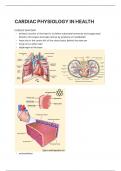
• heart sits in the centre left of the chest cavity behind the sternum
• lungs sit on either side
• diaphragm at the base
• endocardium
, ◦ similar to endothelium of the blood vessels
• myocardium
◦ striated mononucleated cardiomyocytes
◦ rich in mitochondria
• pericardium - 2 layers
◦ fibrous outermost layer attached to great vessels and sometimes to the
diaphragm
◦ serous - has 2 layers - visceral and parietal
◦ space in between is called the pericardial cavity and is filled with pericardial fluid
which acts as a lubricant
• myocardium is branched
◦ this enables the heart to squeeze as opposed to contract in one direction like
skeletal muscle
• striated nucleated cardiomyocytes are rich in mitochondria
◦ lines visible through cells
◦ indicate sarcomere edges
, • 4 cardiac chambers (2 atria & 2 ventricles)
• 4 sets of valves (made of fibrous tissue)
• Tricuspid (b/w rt. Atrium and Ventricle)
• Pulmonary (b/w rt. Ventricle and pulmonary artery
• Mitral (b/w lt. Atrium and Ventricle)
• Aortic (b/w lt. Ventricle and Aorta)
CARDIAC ANATOMY
• primary function of the heart is to deliver substrate hormones and oxygenated
blood to the organs and help remove by products of metabolism
• heart sits in the centre left of the chest cavity behind the sternum
• lungs sit on either side
• diaphragm at the base
• endocardium
, ◦ similar to endothelium of the blood vessels
• myocardium
◦ striated mononucleated cardiomyocytes
◦ rich in mitochondria
• pericardium - 2 layers
◦ fibrous outermost layer attached to great vessels and sometimes to the
diaphragm
◦ serous - has 2 layers - visceral and parietal
◦ space in between is called the pericardial cavity and is filled with pericardial fluid
which acts as a lubricant
• myocardium is branched
◦ this enables the heart to squeeze as opposed to contract in one direction like
skeletal muscle
• striated nucleated cardiomyocytes are rich in mitochondria
◦ lines visible through cells
◦ indicate sarcomere edges
, • 4 cardiac chambers (2 atria & 2 ventricles)
• 4 sets of valves (made of fibrous tissue)
• Tricuspid (b/w rt. Atrium and Ventricle)
• Pulmonary (b/w rt. Ventricle and pulmonary artery
• Mitral (b/w lt. Atrium and Ventricle)
• Aortic (b/w lt. Ventricle and Aorta)