Basal Cell Carcinoma
Definition & DDx DDx
Originating from basal keratinocytes - SCC
usually 2º to DNA damage by UV radiation - Melanoma
- Invasion of basement membrane - Seborrhoeic keratosis
- Most common skin cancer - Actinic keratosis
- Most common western cancer - Dermatofibroma
Classification Anatomy & Risks
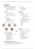
Nodular Risk factors
- Most common subtype - FHx/PMH
- Raised, shiny, pink/translucent - Genetic syndromes e.g., Gorlin
nodule Syndrome
- With central ulceration/crusting o AD loss of tumour
Other suppressor gene function
- Sclerosing PTCH
- Keratotic - Fitzpatrick type I/II skin
- Pigmented (rare) - High level sun/UV exposure e.g.,
- Superficial (often mistaken for occupational, sunburn, sunbeds
eczema/psoriasis - Immunosuppression
- Chronic inflammation e.g., burn
site
- Smoking
- Old age
- Male
Investigations
Diagnosis via excision biopsy with 4mm
margin
- Rx and diagnosis based on clinical
suspicion
Symptoms & Complications - Occasionally, punch biopsy
- Very slow growing completed first
- Local destruction (rare metastasis) 6mm margin for high-risk lesions
- Usually asymptomatic - 2cm diameter
- Central ulceration - Location on
- Sun-exposed areas ear/lip/face/hands/feet/genitals
- Central depression - Immunosuppressed
- Pearly surface/rolled edge - Recurrent disease
- Telangiectasia
Treatment/Management & Side effects
As above; sometimes Mohs micrographic surgery used
- Tissue removed and examined under microscope in real time
Lifestyle advice to prevent further lesions e.g., suncream
,Squamous Cell Carcinoma
Definition & DDx
Locally invasive malignant tumour of Investigations
epidermal keratinocytes Poor prognosis
Risk factors - Poorly differentiated
- Excessive sun exposure - ≥2cm diameter
- AK/Bowen’s disease - >4mm deep
- Immunosuppression e.g., - Immunosuppression
transplant, HIV
- Smoking Treatment/Management & Side effects
- Long-standing leg ulcers Surgical excision
- Genetic conditions e.g., xeroderma - <2cm diameter = 4mm margin
pigmentosum excision
- ≥2cm diameter = 6mm margin
Symptoms & Complications excision
- Sun exposed sites Mohs micrographic surgery may be used in
- Rapidly expanding; may bleed high-risk patients and cosmetically
- Painless/Ulcerated nodules important sites
- ‘Cauliflower-like’ appearance AK Rx = 5-fluorouracil/NSAIDS
Malignant Melanoma
Definition & DDx Anatomy & Risks
Skin cancer arising from melanocytes Risk factors
Subtypes - FHx/PMH
- Superficial spreading - Genetic syndromes
- Lentigo maligna - Fitzpatrick I/II
- Acral lentiginous (involves - Immunosuppression
palms/soles) - High levels of sun/UV
- Subungual - Presence of atypical melanocytic
- Amelanotic naevi
Symptoms & Complications - Smoking
ABCDE - Advanced age
- Asymmetry - Male
- Border irregularity Investigations
- Colour variation - Check for distant metastases
- Diameter ≥7mm o PET/CT may be necessary
- Elevation/evolution over time - Excisional biopsy with 2mm margin
Can also include itchiness and bleeding - If Breslow thickness >1mm =
sentinel LN biopsy
Treatment/Management & Side effects
Breslow thickness determined histologically = most important prognostic indicator
- Mm from top of granular layer to deepest point of tumour
- Breslow thickness in mm x10 = further excision outside 2mm margin required
May also require adjuvant immunotherapy/chemotherapy in mets/Stage III/IV
- Pembrolizumab
50% 5-yr survival if Breslow thickness ≥4mm:
- Best prognosis in <0.75mm
, Pressure Sores
Anatomy & Risks
Develop in pts unable to move parts of body due to illness, paralysis or advancing age
Risk factors
- Malnourishment
- Incontinence
- Lack of mobility
- Pain (leading to reduction in mobility)
Symptoms & Complications
Waterlow score (screening); includes:
- BMI
- Nutrition status
- Skin type
- Mobility
- Continence
Investigations
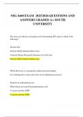
Grading 1-4
I. Non-blanchable erythema of intact skin
II. Partial thickness skin loss involving epidermis/dermis; superficial abrasion/blister
III. Full thickness skin loss; damage/necrosis of SC tissue; not through fascia
IV. Extensive destruction/necrosis/damage to muscle/bone/structures +/- full
thickness skin loss
Treatment/Management & Side effects
- Moist wound environment encourages ulcer healing
o Hydrocolloid dressings/hydrogels
o Avoid soap to stop drying out
- Only use abx if evidence of surrounding cellulitis
- Consider referral to tissue viability nurse
- Surgical debridement may be beneficial for selected wounds
Other skin lesions
Seborrhoeic keratosis
‘Stuck on’ warty plaque with fissured keratin surface
Melanocytic naevi
Can be junctional/compound/intradermal
Dermatofibroma
Small firm nodule <1cm; slow growth; asymptomatic
Definition & DDx DDx
Originating from basal keratinocytes - SCC
usually 2º to DNA damage by UV radiation - Melanoma
- Invasion of basement membrane - Seborrhoeic keratosis
- Most common skin cancer - Actinic keratosis
- Most common western cancer - Dermatofibroma
Classification Anatomy & Risks
Nodular Risk factors
- Most common subtype - FHx/PMH
- Raised, shiny, pink/translucent - Genetic syndromes e.g., Gorlin
nodule Syndrome
- With central ulceration/crusting o AD loss of tumour
Other suppressor gene function
- Sclerosing PTCH
- Keratotic - Fitzpatrick type I/II skin
- Pigmented (rare) - High level sun/UV exposure e.g.,
- Superficial (often mistaken for occupational, sunburn, sunbeds
eczema/psoriasis - Immunosuppression
- Chronic inflammation e.g., burn
site
- Smoking
- Old age
- Male
Investigations
Diagnosis via excision biopsy with 4mm
margin
- Rx and diagnosis based on clinical
suspicion
Symptoms & Complications - Occasionally, punch biopsy
- Very slow growing completed first
- Local destruction (rare metastasis) 6mm margin for high-risk lesions
- Usually asymptomatic - 2cm diameter
- Central ulceration - Location on
- Sun-exposed areas ear/lip/face/hands/feet/genitals
- Central depression - Immunosuppressed
- Pearly surface/rolled edge - Recurrent disease
- Telangiectasia
Treatment/Management & Side effects
As above; sometimes Mohs micrographic surgery used
- Tissue removed and examined under microscope in real time
Lifestyle advice to prevent further lesions e.g., suncream
,Squamous Cell Carcinoma
Definition & DDx
Locally invasive malignant tumour of Investigations
epidermal keratinocytes Poor prognosis
Risk factors - Poorly differentiated
- Excessive sun exposure - ≥2cm diameter
- AK/Bowen’s disease - >4mm deep
- Immunosuppression e.g., - Immunosuppression
transplant, HIV
- Smoking Treatment/Management & Side effects
- Long-standing leg ulcers Surgical excision
- Genetic conditions e.g., xeroderma - <2cm diameter = 4mm margin
pigmentosum excision
- ≥2cm diameter = 6mm margin
Symptoms & Complications excision
- Sun exposed sites Mohs micrographic surgery may be used in
- Rapidly expanding; may bleed high-risk patients and cosmetically
- Painless/Ulcerated nodules important sites
- ‘Cauliflower-like’ appearance AK Rx = 5-fluorouracil/NSAIDS
Malignant Melanoma
Definition & DDx Anatomy & Risks
Skin cancer arising from melanocytes Risk factors
Subtypes - FHx/PMH
- Superficial spreading - Genetic syndromes
- Lentigo maligna - Fitzpatrick I/II
- Acral lentiginous (involves - Immunosuppression
palms/soles) - High levels of sun/UV
- Subungual - Presence of atypical melanocytic
- Amelanotic naevi
Symptoms & Complications - Smoking
ABCDE - Advanced age
- Asymmetry - Male
- Border irregularity Investigations
- Colour variation - Check for distant metastases
- Diameter ≥7mm o PET/CT may be necessary
- Elevation/evolution over time - Excisional biopsy with 2mm margin
Can also include itchiness and bleeding - If Breslow thickness >1mm =
sentinel LN biopsy
Treatment/Management & Side effects
Breslow thickness determined histologically = most important prognostic indicator
- Mm from top of granular layer to deepest point of tumour
- Breslow thickness in mm x10 = further excision outside 2mm margin required
May also require adjuvant immunotherapy/chemotherapy in mets/Stage III/IV
- Pembrolizumab
50% 5-yr survival if Breslow thickness ≥4mm:
- Best prognosis in <0.75mm
, Pressure Sores
Anatomy & Risks
Develop in pts unable to move parts of body due to illness, paralysis or advancing age
Risk factors
- Malnourishment
- Incontinence
- Lack of mobility
- Pain (leading to reduction in mobility)
Symptoms & Complications
Waterlow score (screening); includes:
- BMI
- Nutrition status
- Skin type
- Mobility
- Continence
Investigations
Grading 1-4
I. Non-blanchable erythema of intact skin
II. Partial thickness skin loss involving epidermis/dermis; superficial abrasion/blister
III. Full thickness skin loss; damage/necrosis of SC tissue; not through fascia
IV. Extensive destruction/necrosis/damage to muscle/bone/structures +/- full
thickness skin loss
Treatment/Management & Side effects
- Moist wound environment encourages ulcer healing
o Hydrocolloid dressings/hydrogels
o Avoid soap to stop drying out
- Only use abx if evidence of surrounding cellulitis
- Consider referral to tissue viability nurse
- Surgical debridement may be beneficial for selected wounds
Other skin lesions
Seborrhoeic keratosis
‘Stuck on’ warty plaque with fissured keratin surface
Melanocytic naevi
Can be junctional/compound/intradermal
Dermatofibroma
Small firm nodule <1cm; slow growth; asymptomatic