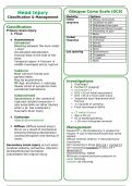
Head Injury Glasgow Coma Scale (GCS)
Classification & Management Modality Options
Motor 6 Obeys commands
response 5 Localises to pain
4 Withdraws from pain
Classification 3 Abnormal flexion to pain
Primary brain injury 2 Extending to pain
1. Focal 1 None
Verbal 5 Orientated
Haematomas response 4 Confused
Extradural 3 Words
Bleeding between the dura mater 2 Sounds
+ skull 1 None
Acceleration-deceleration Eye opening 4 Spontaneous
trauma/ blow to the side of the 3 To speech
head 2 To pain
Temporal region fracture 1 None
middle meningeal artery rupture
Subdural
Most common frontal and
parietal lobes
May be acute/chronic Investigations
RF Age, alcoholism + anticoag 1.Discharge?
Slower onset of symptoms than 2.Further CT imaging?
extradural haematoma a.Immediate CT
GCS <13 on initial assessment
Subarachnoid GCS <15 at 2 hours post-injury
Spontaneous in the context of Suspected open /depressed skull
ruptured cerebral aneurysm + fracture
also association w/ other injuries >1 episode of vomiting
when a pt. has sustained a Focal neurological deficit
traumatic brain injury
b. CT within 8 hours
Age ≥ 65 yrs
Contusion Hx of bleeding/clotting
adjacent/contralateral > 30 mins retrograde amnesia
2. Diffuse (Diffuse axonal injury) Management
Occur as a result of mechanical Raised ICP + life threatening prepare for
shearing following deceleration, theatre use IV Mannitol/Furosemide whilst
causing disruption and tearing of waiting
axons Diffuse cerebral oedema decompressive
craniotomy
Secondary brain injury occurs when ICP monitoring in those GCS 3-8 + normal CT
cerebral oedema, ischaemia, infection, ICP monitoring mandatory in GCS 3-8 +
tonsillar/tentorial herniation abnormal CT
exacerbates the original injury Hyponatraemia- SIADH
Minimum cerebral perfusion pressure of:
Adult 70mmHg
Children 40-70mmHg