Respiratory System Basics
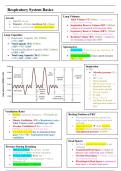
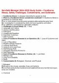
Lung Volumes
Alveoli
• Tidal Volume (VT) 500ml - volume of air
• 300x106 alveoli moving in/out of lungs
• Diameter ~0.2mm, membrane SA ~0.4m
• Inspiration Reserve Volume (IRV) 3000ml -
• Surface covered with moist alveolar lining volume of air inspired with maximal effort
• Expiratory Reserve Volume (ERV) 1200ml
Lung Capacities - volume of air expired with maximal effort
• Inspiratory Capacity (IC) 3500ml • Residual Volume (RV) 1200ml - volume of
= VT + IRV air remaining in lungs after ERV
• Vital Capacity (VC) 4700ml
= IRV + VT + ERV Spirometers
• Functional Residual Capacity (FRC) 2400ml • Inspiration → pen moves up, expiration →
= ERV + RV pen moves down
• Total Lung Capacity (TLC) 5900ml • Cannot measure RV, hence cannot measure
= VT + IRV + ERV + RV FRC or TLC
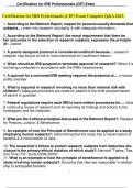
Inspiration
At FRC:
• Alveolar pressure (PA)
=0
• IPP = -ve
1. Chest expands
2. IPP becomes more -ve
3. Increased outward
force across alveoli
4. Alveoli expand
5. PA falls
6. Pmouth > Palveoli
7. Air flows to alveoli
until pressure returns to
zero
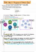
Ventilation Rates
• Resting respiratory frequency ~15 breaths per Resting Position at FRC
min • All respiratory muscles relaxed, but
• Minute Ventilation (VE) = Respiratory rate x lungs recoil inwards and chest recoils
Tidal Volume (total ventilation per min) outwards
• Alveolar Ventilation (VA) = volume of • Opposing recoils → slight negative
FRESH air reaching alveoli per min pressure between pleural membranes
• VA differs from VE due to anatomical dead (intrapleural pressure, PIP/IPP)
space: VA = VE - Anatomical dead space
ventilation
Dead Spaces
• Air that is inhaled but not used in gas
Pressure During Breathing exchange
• Quiet breathing: PIP always -ve, but • Anatomical Dead Space = volume of
inspiration is more -ve than expiration pharynx and conducting zone (150ml)
• Forced expiration: PIP is +ve • Alveolar Dead Space = volume of air in
• PA: inspiration = -ve, expiration = +ve, in non-functional alveoli
between = 0 • Physiological Dead Space = anatomical
• PA always more +ve than PIP dead space + alveolar dead space
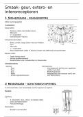
, Respiratory Tract Anatomy
Respiratory Tract Respiratory Epithelium
Upper • Lines much of upper respiratory tract &
• Nasal cavity conducting zone (but NOT alveoli)
• Paranasal sinuses • Pseudostratified, columnar, ciliated
• Nasopharynx epithelium with goblet cells
Lower • Goblet Cells → mucus, which traps dust
• Larynx (aided by submucosal glands)
Conducting Zone
• Trachea – no gas exchange
• Cilia: beat to propel mucus to pharynx
• Bronchi (1/2/3)
• Terminal bronchioles Cilia structure: 20 microtubules (9
• Respiratory bronchioles Respiratory Zone doublets + central pair), 7-10m
• Alveolar ducts –gas exchange
• Alveolar sacs Bronchioles
• Columnar → cuboidal ciliated cells in
respiratory epithelium
Conducting Zone
• Discrete bundles of smooth muscle
From trachea → terminal bronchioles
• No cartilage or submucosal glands + fewer
• Tall columnar → cuboidal epithelium
goblet cells than bronchi
• No smooth muscle → complete smooth
muscle layer → discrete bundles
• Many submucosal glands → none Terminal Bronchioles
• Many goblet cells → none • Cuboidal ciliated respiratory epithelium
• No Clara cells → Clara cells present • No goblet cells, rather Clara cells → secrete
components of surfactant and pumps Cl-
Trachea
• Very tall respiratory epithelium
• Highly cellular/vascular lamina propria + rich
in elastin
• Submucosa contains mucoserous glands
• C-shaped cartilage rings, prevent collapse
• Contraction of trachealis → reduced diameter
→ intrathoracic pressure
Primary Bronchus
• Shorter respiratory epithelium & fewer goblet
cells compared to trachea
• Discontinuous smooth muscle layer secretes
lamina propria & submucosa Respiratory Bronchioles
• No C-shaped rings – plates of cartilage • Minimal gas exchange: single alveoli in
instead walls
• Branch into individual alveoli/alveolar
ducts
Tertiary Bronchus
• Tall, columnar respiratory epithelium with
little pseudostratification Alveolar Ducts/Sacs
• Complete layer of smooth muscle below • Supported by smooth muscle cells, collagen
lamina propria (contraction by PNS) & elastin
• Fewer goblet cells and fewer mucoserous • ~50 alveoli per sac
glands than trachea • Site of most gas exchange with extensive
• Irregular cartilage plates blood supply / capillary network
Lung Volumes
Alveoli
• Tidal Volume (VT) 500ml - volume of air
• 300x106 alveoli moving in/out of lungs
• Diameter ~0.2mm, membrane SA ~0.4m
• Inspiration Reserve Volume (IRV) 3000ml -
• Surface covered with moist alveolar lining volume of air inspired with maximal effort
• Expiratory Reserve Volume (ERV) 1200ml
Lung Capacities - volume of air expired with maximal effort
• Inspiratory Capacity (IC) 3500ml • Residual Volume (RV) 1200ml - volume of
= VT + IRV air remaining in lungs after ERV
• Vital Capacity (VC) 4700ml
= IRV + VT + ERV Spirometers
• Functional Residual Capacity (FRC) 2400ml • Inspiration → pen moves up, expiration →
= ERV + RV pen moves down
• Total Lung Capacity (TLC) 5900ml • Cannot measure RV, hence cannot measure
= VT + IRV + ERV + RV FRC or TLC
Inspiration
At FRC:
• Alveolar pressure (PA)
=0
• IPP = -ve
1. Chest expands
2. IPP becomes more -ve
3. Increased outward
force across alveoli
4. Alveoli expand
5. PA falls
6. Pmouth > Palveoli
7. Air flows to alveoli
until pressure returns to
zero
Ventilation Rates
• Resting respiratory frequency ~15 breaths per Resting Position at FRC
min • All respiratory muscles relaxed, but
• Minute Ventilation (VE) = Respiratory rate x lungs recoil inwards and chest recoils
Tidal Volume (total ventilation per min) outwards
• Alveolar Ventilation (VA) = volume of • Opposing recoils → slight negative
FRESH air reaching alveoli per min pressure between pleural membranes
• VA differs from VE due to anatomical dead (intrapleural pressure, PIP/IPP)
space: VA = VE - Anatomical dead space
ventilation
Dead Spaces
• Air that is inhaled but not used in gas
Pressure During Breathing exchange
• Quiet breathing: PIP always -ve, but • Anatomical Dead Space = volume of
inspiration is more -ve than expiration pharynx and conducting zone (150ml)
• Forced expiration: PIP is +ve • Alveolar Dead Space = volume of air in
• PA: inspiration = -ve, expiration = +ve, in non-functional alveoli
between = 0 • Physiological Dead Space = anatomical
• PA always more +ve than PIP dead space + alveolar dead space
, Respiratory Tract Anatomy
Respiratory Tract Respiratory Epithelium
Upper • Lines much of upper respiratory tract &
• Nasal cavity conducting zone (but NOT alveoli)
• Paranasal sinuses • Pseudostratified, columnar, ciliated
• Nasopharynx epithelium with goblet cells
Lower • Goblet Cells → mucus, which traps dust
• Larynx (aided by submucosal glands)
Conducting Zone
• Trachea – no gas exchange
• Cilia: beat to propel mucus to pharynx
• Bronchi (1/2/3)
• Terminal bronchioles Cilia structure: 20 microtubules (9
• Respiratory bronchioles Respiratory Zone doublets + central pair), 7-10m
• Alveolar ducts –gas exchange
• Alveolar sacs Bronchioles
• Columnar → cuboidal ciliated cells in
respiratory epithelium
Conducting Zone
• Discrete bundles of smooth muscle
From trachea → terminal bronchioles
• No cartilage or submucosal glands + fewer
• Tall columnar → cuboidal epithelium
goblet cells than bronchi
• No smooth muscle → complete smooth
muscle layer → discrete bundles
• Many submucosal glands → none Terminal Bronchioles
• Many goblet cells → none • Cuboidal ciliated respiratory epithelium
• No Clara cells → Clara cells present • No goblet cells, rather Clara cells → secrete
components of surfactant and pumps Cl-
Trachea
• Very tall respiratory epithelium
• Highly cellular/vascular lamina propria + rich
in elastin
• Submucosa contains mucoserous glands
• C-shaped cartilage rings, prevent collapse
• Contraction of trachealis → reduced diameter
→ intrathoracic pressure
Primary Bronchus
• Shorter respiratory epithelium & fewer goblet
cells compared to trachea
• Discontinuous smooth muscle layer secretes
lamina propria & submucosa Respiratory Bronchioles
• No C-shaped rings – plates of cartilage • Minimal gas exchange: single alveoli in
instead walls
• Branch into individual alveoli/alveolar
ducts
Tertiary Bronchus
• Tall, columnar respiratory epithelium with
little pseudostratification Alveolar Ducts/Sacs
• Complete layer of smooth muscle below • Supported by smooth muscle cells, collagen
lamina propria (contraction by PNS) & elastin
• Fewer goblet cells and fewer mucoserous • ~50 alveoli per sac
glands than trachea • Site of most gas exchange with extensive
• Irregular cartilage plates blood supply / capillary network