BIOD151 - Module 5 Summary, Complete
Solution With pictures and diagrams For Your
Module 5 Exams.
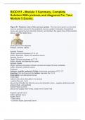
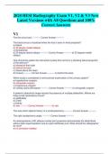
Figure 5.1 Posterior view of the nervous system. The brain and spinal cord (central
nervous system) connect to the peripheral nervous system. Examples of peripheral
nerves are spinal nerves (cervical, thoracic, and lumbar), the upper trunk of the brachial
plexus, and the radial nerve.
(3) Divisions of the Spinalis
thoracis, cervicis, capitis
Spinalis Thoracis
Origin: spinous processes of T11-L2
Action: extension / flexion of vertebral column
Spinalis Cervicis
Origin: Spinous processes of C7-T2
Action: extend and laterally flex spine
Spinalis Capitis
Origin: spinous processes of lower cervical and upper thoracic vertebrae
Action: extends vertebral column
Scalenes
(anterior, middle, posterior) Origin: transverse processes of C2- C7
Insertion: first and second ribs Action: elevates ribs 1 & 2
Innervation: cervical spinal nerves
Innervation
refers to the nerve stimulation of a muscle
Lumbar Plexus (L1-L4)
plexus that includes the femoral nerve and obturator nerve - supplies abdominal wall,
anterior thigh, and genitalia
Sacral Plexus (L4-S4)
plexus that supply lower limbs, sciatic nerve: lower limb
Superior gluteal nerve
Inferior gluteal nerve
Sciatic nerve
• Tibial nerve
• Common fibular (peroneal) nerve
,(4) Plexuses
cervical, brachial, lumbar, sacral
Brachial Plexus (C5-T1)
plexus that includes the axillary nerve, musculocutaneous nerve, radial nerve, median
nerve, ulnar nerve - nerve supply to the upper extremities (spine, shoulder, arm, hand)
Cervical Plexus (C1-C5)
plexus that supplies neck and phrenic nerve to the diaphragm
Accessory Nerve
motor fibers to neck and upper back - CN XI
Motor Actions
messages from the CNS to a muscle
Sensation / Sensory Input
messages received by the CNS from the external environment (figure 5.1 and 5.2)
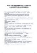
Figure 5.2 Peripheral nerves carry the communication from the central nervous system
(brain and spinal cord) to the muscle. Peripheral nerves also carry information from the
environment to the central nervous system.
Peripheral Nerves
Interconnecting branches of spinal nerves
Surrounded by connective tissue sheaths - carry signals from the CNS (brain and spinal
cord) to a specific muscle destination in order to provide movement
Muscle Communication Pathway
Communication within the body to coordinate movement starts in the brain with a
message that is sent through the spinal cord and eventually attaches to a
muscle. Peripheral nerves carry the signal from the central nervous system (brain
and spinal cord) to a specific muscle destination to provide movement. Messages from
the central nervous system to a muscle are called motor actions. Nerves also carry
information from the external environment to the central nervous system, called
sensation or sensory input (see Figure 5.1 and Figure 5.2). Spinal nerves combine to
form complex networks of peripheral nerves throughout the body.
Muscle Fiber
cell containing thousands of myofibrils
Myofibrils
cylindrical in shape and run the length of the muscle fiber. The light microscope shows
that a __________________has light and dark bands called striations. It is these
bands that cause skeletal muscle to appear striated. Striations of __________________
, are formed by protein myofilaments within contractile units
called sarcomeres (see Figure 5.38)
Sarcomeres
a structural unit of a myofibril in striated muscle, consisting of a dark band and the
nearer half of each adjacent pale band. A __________contains two types of
protein myofilaments (also referred to as filaments). The thick filaments are made up of
a protein called myosin, and the thin filaments are made up of a protein called actin.
As a muscle fiber contracts, the _______________within the myofibrils shorten. When a
_________________shortens, the actin (thin) filaments slide past the myosin (thick)
filaments and approach one another. The movement of actin filaments in relation to
myosin filaments causes the muscle to shorten.
Actin
A globular protein that links into chains, two of which twist helically about each other,
forming microfilaments in muscle and other contractile elements in cells.
Myosin
The contractile protein that makes up the thick filaments of muscle fibers
Myofilaments
The contractile proteins, actin and myosin, of muscle cells
Z Lines
connect parallel bands of thin filaments (actin) - outside of the sarcomere - when a
muscle contraction occurs, these lines move closer together towards the center of the
sarcomere (M line)
M Line
supporting proteins that hold the thick filaments (myosin) together in the H zone - middle
of sarcomere
I Band
(light band) appears light when stained because it only contains thin filaments.
A Band
(dark band) contains thin and thick filaments; however, it stains darker because it
contains the thick filaments
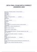
Figure 5.38 View of a muscle fiber, to the microscopic view of a thick and thin
filament. Note the heads on the myosin filaments that enable the work of the muscle
contraction.
Cross Bridges
In the presence of calcium ions, portions of the myosin filaments called ___________-
___________ bend backward and attach to actin filaments. After attaching to the actin
Solution With pictures and diagrams For Your
Module 5 Exams.
Figure 5.1 Posterior view of the nervous system. The brain and spinal cord (central
nervous system) connect to the peripheral nervous system. Examples of peripheral
nerves are spinal nerves (cervical, thoracic, and lumbar), the upper trunk of the brachial
plexus, and the radial nerve.
(3) Divisions of the Spinalis
thoracis, cervicis, capitis
Spinalis Thoracis
Origin: spinous processes of T11-L2
Action: extension / flexion of vertebral column
Spinalis Cervicis
Origin: Spinous processes of C7-T2
Action: extend and laterally flex spine
Spinalis Capitis
Origin: spinous processes of lower cervical and upper thoracic vertebrae
Action: extends vertebral column
Scalenes
(anterior, middle, posterior) Origin: transverse processes of C2- C7
Insertion: first and second ribs Action: elevates ribs 1 & 2
Innervation: cervical spinal nerves
Innervation
refers to the nerve stimulation of a muscle
Lumbar Plexus (L1-L4)
plexus that includes the femoral nerve and obturator nerve - supplies abdominal wall,
anterior thigh, and genitalia
Sacral Plexus (L4-S4)
plexus that supply lower limbs, sciatic nerve: lower limb
Superior gluteal nerve
Inferior gluteal nerve
Sciatic nerve
• Tibial nerve
• Common fibular (peroneal) nerve
,(4) Plexuses
cervical, brachial, lumbar, sacral
Brachial Plexus (C5-T1)
plexus that includes the axillary nerve, musculocutaneous nerve, radial nerve, median
nerve, ulnar nerve - nerve supply to the upper extremities (spine, shoulder, arm, hand)
Cervical Plexus (C1-C5)
plexus that supplies neck and phrenic nerve to the diaphragm
Accessory Nerve
motor fibers to neck and upper back - CN XI
Motor Actions
messages from the CNS to a muscle
Sensation / Sensory Input
messages received by the CNS from the external environment (figure 5.1 and 5.2)
Figure 5.2 Peripheral nerves carry the communication from the central nervous system
(brain and spinal cord) to the muscle. Peripheral nerves also carry information from the
environment to the central nervous system.
Peripheral Nerves
Interconnecting branches of spinal nerves
Surrounded by connective tissue sheaths - carry signals from the CNS (brain and spinal
cord) to a specific muscle destination in order to provide movement
Muscle Communication Pathway
Communication within the body to coordinate movement starts in the brain with a
message that is sent through the spinal cord and eventually attaches to a
muscle. Peripheral nerves carry the signal from the central nervous system (brain
and spinal cord) to a specific muscle destination to provide movement. Messages from
the central nervous system to a muscle are called motor actions. Nerves also carry
information from the external environment to the central nervous system, called
sensation or sensory input (see Figure 5.1 and Figure 5.2). Spinal nerves combine to
form complex networks of peripheral nerves throughout the body.
Muscle Fiber
cell containing thousands of myofibrils
Myofibrils
cylindrical in shape and run the length of the muscle fiber. The light microscope shows
that a __________________has light and dark bands called striations. It is these
bands that cause skeletal muscle to appear striated. Striations of __________________
, are formed by protein myofilaments within contractile units
called sarcomeres (see Figure 5.38)
Sarcomeres
a structural unit of a myofibril in striated muscle, consisting of a dark band and the
nearer half of each adjacent pale band. A __________contains two types of
protein myofilaments (also referred to as filaments). The thick filaments are made up of
a protein called myosin, and the thin filaments are made up of a protein called actin.
As a muscle fiber contracts, the _______________within the myofibrils shorten. When a
_________________shortens, the actin (thin) filaments slide past the myosin (thick)
filaments and approach one another. The movement of actin filaments in relation to
myosin filaments causes the muscle to shorten.
Actin
A globular protein that links into chains, two of which twist helically about each other,
forming microfilaments in muscle and other contractile elements in cells.
Myosin
The contractile protein that makes up the thick filaments of muscle fibers
Myofilaments
The contractile proteins, actin and myosin, of muscle cells
Z Lines
connect parallel bands of thin filaments (actin) - outside of the sarcomere - when a
muscle contraction occurs, these lines move closer together towards the center of the
sarcomere (M line)
M Line
supporting proteins that hold the thick filaments (myosin) together in the H zone - middle
of sarcomere
I Band
(light band) appears light when stained because it only contains thin filaments.
A Band
(dark band) contains thin and thick filaments; however, it stains darker because it
contains the thick filaments
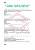
Figure 5.38 View of a muscle fiber, to the microscopic view of a thick and thin
filament. Note the heads on the myosin filaments that enable the work of the muscle
contraction.
Cross Bridges
In the presence of calcium ions, portions of the myosin filaments called ___________-
___________ bend backward and attach to actin filaments. After attaching to the actin