SMOOTH & SKELETAL MUSCLE
SYSTEMS PHYSIOLOGY- MUSCLE
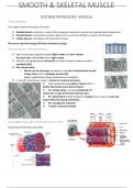
TYPES OF MUSCLE
Our bodies contain three types of muscle:
1) Skeletal Muscle- voluntary – control them to govern movement, posture and regulate body temperature
2) Smooth Muscle- involuntary and lines organs (such as stomach, bladder as well as blood vessels
3) Cardiac Muscle- involuntary and found only in heart
All convert chemical energy (ATP) into mechanical energy
SKELETAL MUSCLE- STRIATED MUSCLE
❖ Light microscope originally revealed light I bands and dark A bands
Fine dark lines called Z-lines bisect light I bands
❖ Electron micrograph shows nucleus (N) in similar location as above as well as
myofibrils (My)
❖ Mt= mitochondria
o Muscle use lot of energy- we expect to see lots of mitochondria as well
energy stores such as glycogen granules (G)
Higher magnification allows visualisation of myofibrils
❖ S- smooth membranous system- involved in muscle contraction
❖ Dark band bisected by lighter H band which is
further bisected by more dense M band
❖ Td- Tubular triads- containing flattened of T
system (T) and pair of terminal cisternae (TC)
❖ M- mitochondria mostly localised in I bands close to parts of actin and
myosin filaments during contraction
❖ SR- sarcoplasmic reticulum
❖ T-tubule- transverse tubule
SARCOMERE- BASIC UNIT OF STRIATED MUSCLE TISSUE
Repeating unit between two Z lines
, SLIDING FILAMENT THEORY
Describes mechanism of muscle contraction
❖ Thick and thin filaments slide over each one another
o Neither of thick or thin filaments shorten
❖ During contractions- H-zone becomes narrower
❖ Elastic titin filaments keep thick filament in central
position
STRUCTURE AND ARRANGEMENT OF MYOSIN MOLECULES WITHIN THICK FILAMENT
❖ Tropomyosin- protein that binds and stabilises actin filaments in cell
o In skeletal and cardiac muscle cells- tropomyosin is released after interacting with troponin and
calcium- facilitating binding of actin to myosin- causes muscle contraction
SYSTEMS PHYSIOLOGY- MUSCLE
TYPES OF MUSCLE
Our bodies contain three types of muscle:
1) Skeletal Muscle- voluntary – control them to govern movement, posture and regulate body temperature
2) Smooth Muscle- involuntary and lines organs (such as stomach, bladder as well as blood vessels
3) Cardiac Muscle- involuntary and found only in heart
All convert chemical energy (ATP) into mechanical energy
SKELETAL MUSCLE- STRIATED MUSCLE
❖ Light microscope originally revealed light I bands and dark A bands
Fine dark lines called Z-lines bisect light I bands
❖ Electron micrograph shows nucleus (N) in similar location as above as well as
myofibrils (My)
❖ Mt= mitochondria
o Muscle use lot of energy- we expect to see lots of mitochondria as well
energy stores such as glycogen granules (G)
Higher magnification allows visualisation of myofibrils
❖ S- smooth membranous system- involved in muscle contraction
❖ Dark band bisected by lighter H band which is
further bisected by more dense M band
❖ Td- Tubular triads- containing flattened of T
system (T) and pair of terminal cisternae (TC)
❖ M- mitochondria mostly localised in I bands close to parts of actin and
myosin filaments during contraction
❖ SR- sarcoplasmic reticulum
❖ T-tubule- transverse tubule
SARCOMERE- BASIC UNIT OF STRIATED MUSCLE TISSUE
Repeating unit between two Z lines
, SLIDING FILAMENT THEORY
Describes mechanism of muscle contraction
❖ Thick and thin filaments slide over each one another
o Neither of thick or thin filaments shorten
❖ During contractions- H-zone becomes narrower
❖ Elastic titin filaments keep thick filament in central
position
STRUCTURE AND ARRANGEMENT OF MYOSIN MOLECULES WITHIN THICK FILAMENT
❖ Tropomyosin- protein that binds and stabilises actin filaments in cell
o In skeletal and cardiac muscle cells- tropomyosin is released after interacting with troponin and
calcium- facilitating binding of actin to myosin- causes muscle contraction