Neural networks: stroke rehabilitation
General introduction
Incidentie
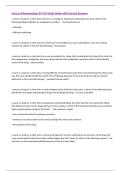
- With increasing age, stroke contributes the most to life years lost by death or disability…
- DALY (Disability-adjusted life years)
Total burden associated with disease (not only mortality)
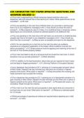
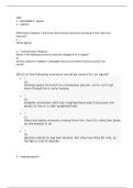
Incdence hemorrhagic stroke (hypertensive)
Pathofysioogie stroke
Ischemic stroke
• Brain requires constant supply of oxygen and glucose, provided by blood
Nala Melis Pagina 1
, Neural networks: stroke rehabilitation
• At rest, brain requires 20 % of total body oxygen consumption
• If blood flow reduces
- To zero: death of brain tissue (infarction) within 4-10 min
- <16-18 ml/100g brain tissue: infarction within 1 hour
- <20-100 ml/ 100g brain tissue: ischemia without infarction, unless persisting for several hours or
day ..
• Pathophysiological cascade
- Due to loss of blood flow
depletion of energy stores (ATP)
leads to ionic imbalances
Leads to electrical disturbances in membrane potential
Cytotoxic edema (extracellular water passes into cells, resulting in their swellings)
- => further cascade of ischemia-related changes:
increase the production of reactive oxygen species (ROS) and nitric oxide (NO).
=> destroys cell membranes, cell lysis, and cell death through mechanisms such as necrosis or
apoptosis
• Infarction region => irreversible damage
• Penumbra region => no immediate cell death, potential for recovery, if early reperfusion is achieved
Two-sided role of Microglia in Penumbra in hersenen
- Role in Neuroinflammation
- Activated in the affected ischemic area and migrate to the penumbra
- Activation peaks 48 to 72 hours after the stroke onset and can persist for several weeks
- Pro-inflammation: production of proinflammatory cytokines such as ROS, NO, interleukin-1β, and
tumor necrosis factor-α => contribute to further death of neurons
- Anti-inflammation : release anti-inflammatory cytokines and neurotrophic factors, including brain-
derived neurotrophic factor, glial cell-line-derived neurotrophic factor, and basic fibroblast growth
factor
=> Participate in cellular debris clearance and tissue repair
Hemorrhagic stroke
Rupture of brain artery (often due to chronic hypertensia)
- => Explosive entry of blood (hematoma) in brain parenchyma
- Meeste door hypertensia (ook nog andere oorzaken)
Primary injury mechanism
- Expanding hematoma volume
Nala Melis Pagina 2
, Neural networks: stroke rehabilitation
Increase intracranial pressure
Often reduced cerebral perfusion
Cause of ischemic injury (cfr. ischemic stroke)
Secondary injury mechanisms
- Lysis of red blood cells, rendering hemoglobin, iron and bilirubin in extracellular space => induction
of inflammatory mechanisms => disruption of BBB (blood bain barrier) =>Perihematomal edema
influx H2O in hersenen (can lead to life-threatening brain edema)
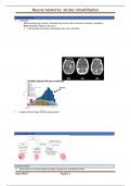
Neuro-imaging of stroke lesion
(hyper)acute
Neuroimaging is a critical component of stroke diagnosis / management
Two main modalities
- Computer tomography (CT) scan (non-contrast)
- MRI neuroimaging
Important for differential diagnosis between
- Ischemic
- Hemorrhagic stroke !!
1. Computed tomography (CT)
Most common type of imaging of acute stroke
It is fast, inexpensive and readily available
- use of X-rays (iets invasiever)
- different tissues interact with X-rays in different ways
- The extent to which a material can be penetrated by an X- ray beam is described in
terms of an absorption/ attenuation coefficient
= assesses how much a beam is weakened by passing through a voxel of tissue
(voxel = volumetric pixel).
Voxel= 3D pixel
- Some tissues will allow the passage of X-rays without influencing them much e.g.
air, does not absorb any X-rays, therefore appears completely black…
- Higher attenuation of beam (in Houndsfield units), “more bright” on CT scan (= hyperdense)
2. Ischemic stroke
2.1. Immediate CT scanning - within 1 hour
Main goal => Direct visualization of the clot or embolism
- Artery with clot becomes hyperdense (brighter) on CT scan
Normal blood: about 40 HU
Clot typically around 100 HU
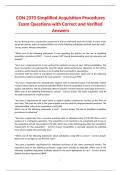
2.2. 1 to 3 hours post stroke
Visualisation of ischemic surrounding tissue
- Grey matter becomes hypodense (darker)
- combination of reduced blood volume and cytotoxic edema (extracellular water held in cells, causing
their swelling)
Few hours post stroke Few days post stroke
2.3. MRI scanning using – Diffusion weighted imaging (DWI = DTI)
Nala Melis Pagina 3
General introduction
Incidentie
- With increasing age, stroke contributes the most to life years lost by death or disability…
- DALY (Disability-adjusted life years)
Total burden associated with disease (not only mortality)
Incdence hemorrhagic stroke (hypertensive)
Pathofysioogie stroke
Ischemic stroke
• Brain requires constant supply of oxygen and glucose, provided by blood
Nala Melis Pagina 1
, Neural networks: stroke rehabilitation
• At rest, brain requires 20 % of total body oxygen consumption
• If blood flow reduces
- To zero: death of brain tissue (infarction) within 4-10 min
- <16-18 ml/100g brain tissue: infarction within 1 hour
- <20-100 ml/ 100g brain tissue: ischemia without infarction, unless persisting for several hours or
day ..
• Pathophysiological cascade
- Due to loss of blood flow
depletion of energy stores (ATP)
leads to ionic imbalances
Leads to electrical disturbances in membrane potential
Cytotoxic edema (extracellular water passes into cells, resulting in their swellings)
- => further cascade of ischemia-related changes:
increase the production of reactive oxygen species (ROS) and nitric oxide (NO).
=> destroys cell membranes, cell lysis, and cell death through mechanisms such as necrosis or
apoptosis
• Infarction region => irreversible damage
• Penumbra region => no immediate cell death, potential for recovery, if early reperfusion is achieved
Two-sided role of Microglia in Penumbra in hersenen
- Role in Neuroinflammation
- Activated in the affected ischemic area and migrate to the penumbra
- Activation peaks 48 to 72 hours after the stroke onset and can persist for several weeks
- Pro-inflammation: production of proinflammatory cytokines such as ROS, NO, interleukin-1β, and
tumor necrosis factor-α => contribute to further death of neurons
- Anti-inflammation : release anti-inflammatory cytokines and neurotrophic factors, including brain-
derived neurotrophic factor, glial cell-line-derived neurotrophic factor, and basic fibroblast growth
factor
=> Participate in cellular debris clearance and tissue repair
Hemorrhagic stroke
Rupture of brain artery (often due to chronic hypertensia)
- => Explosive entry of blood (hematoma) in brain parenchyma
- Meeste door hypertensia (ook nog andere oorzaken)
Primary injury mechanism
- Expanding hematoma volume
Nala Melis Pagina 2
, Neural networks: stroke rehabilitation
Increase intracranial pressure
Often reduced cerebral perfusion
Cause of ischemic injury (cfr. ischemic stroke)
Secondary injury mechanisms
- Lysis of red blood cells, rendering hemoglobin, iron and bilirubin in extracellular space => induction
of inflammatory mechanisms => disruption of BBB (blood bain barrier) =>Perihematomal edema
influx H2O in hersenen (can lead to life-threatening brain edema)
Neuro-imaging of stroke lesion
(hyper)acute
Neuroimaging is a critical component of stroke diagnosis / management
Two main modalities
- Computer tomography (CT) scan (non-contrast)
- MRI neuroimaging
Important for differential diagnosis between
- Ischemic
- Hemorrhagic stroke !!
1. Computed tomography (CT)
Most common type of imaging of acute stroke
It is fast, inexpensive and readily available
- use of X-rays (iets invasiever)
- different tissues interact with X-rays in different ways
- The extent to which a material can be penetrated by an X- ray beam is described in
terms of an absorption/ attenuation coefficient
= assesses how much a beam is weakened by passing through a voxel of tissue
(voxel = volumetric pixel).
Voxel= 3D pixel
- Some tissues will allow the passage of X-rays without influencing them much e.g.
air, does not absorb any X-rays, therefore appears completely black…
- Higher attenuation of beam (in Houndsfield units), “more bright” on CT scan (= hyperdense)
2. Ischemic stroke
2.1. Immediate CT scanning - within 1 hour
Main goal => Direct visualization of the clot or embolism
- Artery with clot becomes hyperdense (brighter) on CT scan
Normal blood: about 40 HU
Clot typically around 100 HU
2.2. 1 to 3 hours post stroke
Visualisation of ischemic surrounding tissue
- Grey matter becomes hypodense (darker)
- combination of reduced blood volume and cytotoxic edema (extracellular water held in cells, causing
their swelling)
Few hours post stroke Few days post stroke
2.3. MRI scanning using – Diffusion weighted imaging (DWI = DTI)
Nala Melis Pagina 3