Cell Cycle
Cell Cycle
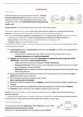
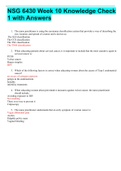
Cell reproduction occurs via elaborate series of events – Cell Cycle
Cell Cycle (Cell-Division Cell)- Reproductive cycle of cell- orderly
sequence of events by which cell duplicates its chromosomes and
usually other contents. And DIVIDES INTO TWO Genetically identical
daughter cells
During interphase- cell is actively expressing its genes and is synthesising proteins.
Chromosome duplication occurs during S phase (S for DNA synthesis)- produce two closely paired sister DNA
molecules - requires 10-12 hrs and occupies about half of cell cycle in typical mammalian cell
After S phase- chromosome segregation and cell division occur in M phase (M for Mitosis)- requires much less time
- comprised of two major events- nuclear division (mitosis) and cytoplasmic division (Cytokinesis)
- Nuclear division- copied chromosomes were distributed into pair of daughter nuclei
- Cytoplasmic division- cells itself divides into two
At end of S phase- DNA molecules in each pair of duplicated chromosomes are intertwined and held together tightly
by specialised protein linkages
Long Interphase- genes are expressed and chromosomes are replicated- two replicas remaining together as
pair of sister chromatids
o chromosomes are extended and much of their chromatin exists as long threads in nucleus-
individual chromosomes can’t easily be distinguished
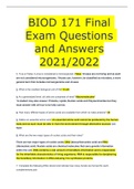
Prophase- two DNA molecules are gradually disentangled and condenses into pairs of rigid rods- SISTER
CHROMATIDS- remain linked by sister chromatid cohesion
o Highly condensed chromosomes in dividing cell- mitotic chromosomes
o Presence of centromere- allows one copy of each duplicated and condensed chromosomes to pulled
into each daughter cell when cell divides
o Kinetochore forms at centromere and attaches duplicated chromosomes to be mitotic spindle- allow
them to be pulled apart
When nuclear envelope disassembles later in mitosis- sister-chromatid pairs become attached to mitotic
spindle (giant bipolar array of molecules)
Siter chromatids are attached to opposite poled of spindle
Align at spindle equator in Metaphase
Destruction of sister-chromatids at start of Anaphase- separated sister chromatids- pulled to opposite poles
of spindle
Spindle is disassembles and segregated chromosome are packaged into separate nuclei at Telophase
Cytokinesis- cleaves cell into two- each daughter cell inherits one of two nuclei
Telomeres- ends of chromosome. Contain repeated nucleotide sequence- enables ends of chromosomes to be
efficiently replicated
- repeated telomere DNA sequences- together with regions adjoining them- form structures that protect ends of
chromosomes from being mistaken by cell for broken DNA molecule in need of repair
Cell Reproduction
Cell reproduction begins with duplication of cell’s components- includes exact
duplication of each chromosome in S PHASE.
Components are divided equally between two daughter cells in M PHASE
,Cell organisation and genome architecture DIFFER DRAMATICALLY between interphase and mitotic cells
DNA in eukaryotic cells packaged in chromatin
DNA of chromosome is packaged in variety of protein components- Histones and various regulatory proteins
involved in control of gene expression
Duplication of chromosome requires replication of DNA and duplication of
chromatin proteins and their proper assembly of DNA
Chromatin- complex of histones, non-histone proteins and nuclear DNA
- allows for DNA compaction and is involved in regulation of all DNA
activities
Production do chromatin increases during S phase- provide raw material needed to package newly synthesised
DNA
- S-Cdks- stimulate large increase in synthesis of four histone subunits that form histone octamers at core of each
nucleosome
Chromatin packing helps to control gene expression:
Heterochromatin- chromatin highly condensed
Activity of genes is modified or suppressed
Euchromatin- more open structures
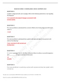
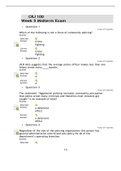
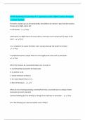
Nucleosomes are basic structural units of chromatin
Nucleases- isolate nucleosomes form chromatin by digestion- break down DNA by cutting
between nucleosome
- Exposed DNA between nucleosome core particles, linker DNA is degraded
Histone octamer- formed from dimers- H3-H4 and H2A- H2B- each in two copies
Histones are small, highly basic proteins- Each of core histones (from octamer) possesses two functional
domains:
- N-Amino-terminal tail- extends out form DNA histone core
- Histone fold- formed from three α-helices- connected by two loops
Histone folds interact with each other allowing formation of dimers- via ‘handshake’ interaction
Histones are among most highly conserved eukaryotic proteins- e.g. amino acid sequence of histone H4
from pea differs from that of cow only 2 of 102 positions
Strong evolutionary conservation- suggests functions of histones involve nearly all of their amino acids-
change in any position is deleterious to cell
Histone folds first bind to each other- form H3-H4 and H2A-H2B dimers and H3-H4 dimers combine to form
tetramers
H3-H4 tetramer further combines with two H2A-H2B dimers- form compact octamer- around which DNA is
wound
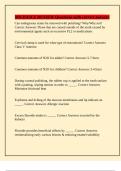
Nucleosomes- composed of DNA and proteins- Histones
147 Base Pairs of double helix DNA winds around histone octamer- like thread around spool- forms slightly
less than two turns
Linker Histones (H1)- bind both DNA and nucleosome core (each nucleosome)
- Can change path of DNA that exists nucleosome – affects linker
DNA accessibility, organisation and higher order chromatin fibre and chromatic
, compaction
Presence of many other DNA-binding proteins, as well as proteins that bind directly to histone- add
important additional features to any array of nucleosomes
Higher order organisation of chromatin
Currently its unknown how exactly 30nm fibre forms
Models explain it’s structures e.g. ‘zig-zag’- likely that histone H1
participates in formation of higher order chromatin structures
In interphase- nuclei chromatin is arranged in loops. Architectural proteins involved in formation of these
loops
Loops of chromatin- compacted by further folding. Genes contained in loop are expressed, loop unfold
and allows cell’s machinery access to DNA
To components identified so far:
- Protein complex- COHESIN and CTCF
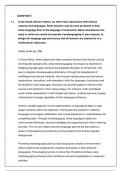
Further hierarchical organisation of interphase chromatin:
- Groups of chromatin loops form Topologically
Associating Domains (TADs)
- TADs are grouped into compartments- compartments may be transcriptionally active (Type A) or
inactive (Type B)
- Compartments belong to individual Chromosomal Territories- occupied by single chromosomes
decondensed after mitosis
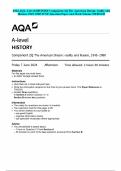
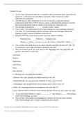
Chromosomal Territories can be visualised using Fluorescent Staining
Technique to paint chromosomes using multi-colour FISH- Spectral Karyotyping- helps
visualise entire chromosomes both in mitosis and during interphase
Interphase chromatin- Conclusions
1. In eukaryotic chromatin DNA is wrapped around histone octamers- nucleosomes
2. During Interphase- chromatin fibres are arranged in loops
Cohesins and CTCF proteins- define boundaries of most of these loops
3. Looping of chromatin fibres has function in chromatin compaction and in regulation
of gene expression
4. Chromatin loop are organised in Topologically Associating Domains (TADs)
5. TADs are organised into compartments- can be transcriptionally active/ inactive
6. Compartments form Chromosome Territories within interphase nuclei
7. Position of gene within these structures affects activity of that gene
8. Genome Organisation- depends on organism, cell type (Tissue), stage of
development, cell cycle phase, current physiological status (e.g. stimuli from environment, stress) and is
disturbed in many pathological states
Structure of mitotic chromosomes
At end of S-phase, long DNA molecules of sister chromatids are tangles in mass of partially
catenated DNA and proteins
Any attempt to pull sisters apart in this state- lead to breaks in chromosome. To avoid this-
sister chromatids reorganise into short and distinct structures- that can be pulled apart
more easily in Anaphase.
These conformation changes involve:
Cell Cycle
Cell reproduction occurs via elaborate series of events – Cell Cycle
Cell Cycle (Cell-Division Cell)- Reproductive cycle of cell- orderly
sequence of events by which cell duplicates its chromosomes and
usually other contents. And DIVIDES INTO TWO Genetically identical
daughter cells
During interphase- cell is actively expressing its genes and is synthesising proteins.
Chromosome duplication occurs during S phase (S for DNA synthesis)- produce two closely paired sister DNA
molecules - requires 10-12 hrs and occupies about half of cell cycle in typical mammalian cell
After S phase- chromosome segregation and cell division occur in M phase (M for Mitosis)- requires much less time
- comprised of two major events- nuclear division (mitosis) and cytoplasmic division (Cytokinesis)
- Nuclear division- copied chromosomes were distributed into pair of daughter nuclei
- Cytoplasmic division- cells itself divides into two
At end of S phase- DNA molecules in each pair of duplicated chromosomes are intertwined and held together tightly
by specialised protein linkages
Long Interphase- genes are expressed and chromosomes are replicated- two replicas remaining together as
pair of sister chromatids
o chromosomes are extended and much of their chromatin exists as long threads in nucleus-
individual chromosomes can’t easily be distinguished
Prophase- two DNA molecules are gradually disentangled and condenses into pairs of rigid rods- SISTER
CHROMATIDS- remain linked by sister chromatid cohesion
o Highly condensed chromosomes in dividing cell- mitotic chromosomes
o Presence of centromere- allows one copy of each duplicated and condensed chromosomes to pulled
into each daughter cell when cell divides
o Kinetochore forms at centromere and attaches duplicated chromosomes to be mitotic spindle- allow
them to be pulled apart
When nuclear envelope disassembles later in mitosis- sister-chromatid pairs become attached to mitotic
spindle (giant bipolar array of molecules)
Siter chromatids are attached to opposite poled of spindle
Align at spindle equator in Metaphase
Destruction of sister-chromatids at start of Anaphase- separated sister chromatids- pulled to opposite poles
of spindle
Spindle is disassembles and segregated chromosome are packaged into separate nuclei at Telophase
Cytokinesis- cleaves cell into two- each daughter cell inherits one of two nuclei
Telomeres- ends of chromosome. Contain repeated nucleotide sequence- enables ends of chromosomes to be
efficiently replicated
- repeated telomere DNA sequences- together with regions adjoining them- form structures that protect ends of
chromosomes from being mistaken by cell for broken DNA molecule in need of repair
Cell Reproduction
Cell reproduction begins with duplication of cell’s components- includes exact
duplication of each chromosome in S PHASE.
Components are divided equally between two daughter cells in M PHASE
,Cell organisation and genome architecture DIFFER DRAMATICALLY between interphase and mitotic cells
DNA in eukaryotic cells packaged in chromatin
DNA of chromosome is packaged in variety of protein components- Histones and various regulatory proteins
involved in control of gene expression
Duplication of chromosome requires replication of DNA and duplication of
chromatin proteins and their proper assembly of DNA
Chromatin- complex of histones, non-histone proteins and nuclear DNA
- allows for DNA compaction and is involved in regulation of all DNA
activities
Production do chromatin increases during S phase- provide raw material needed to package newly synthesised
DNA
- S-Cdks- stimulate large increase in synthesis of four histone subunits that form histone octamers at core of each
nucleosome
Chromatin packing helps to control gene expression:
Heterochromatin- chromatin highly condensed
Activity of genes is modified or suppressed
Euchromatin- more open structures
Nucleosomes are basic structural units of chromatin
Nucleases- isolate nucleosomes form chromatin by digestion- break down DNA by cutting
between nucleosome
- Exposed DNA between nucleosome core particles, linker DNA is degraded
Histone octamer- formed from dimers- H3-H4 and H2A- H2B- each in two copies
Histones are small, highly basic proteins- Each of core histones (from octamer) possesses two functional
domains:
- N-Amino-terminal tail- extends out form DNA histone core
- Histone fold- formed from three α-helices- connected by two loops
Histone folds interact with each other allowing formation of dimers- via ‘handshake’ interaction
Histones are among most highly conserved eukaryotic proteins- e.g. amino acid sequence of histone H4
from pea differs from that of cow only 2 of 102 positions
Strong evolutionary conservation- suggests functions of histones involve nearly all of their amino acids-
change in any position is deleterious to cell
Histone folds first bind to each other- form H3-H4 and H2A-H2B dimers and H3-H4 dimers combine to form
tetramers
H3-H4 tetramer further combines with two H2A-H2B dimers- form compact octamer- around which DNA is
wound
Nucleosomes- composed of DNA and proteins- Histones
147 Base Pairs of double helix DNA winds around histone octamer- like thread around spool- forms slightly
less than two turns
Linker Histones (H1)- bind both DNA and nucleosome core (each nucleosome)
- Can change path of DNA that exists nucleosome – affects linker
DNA accessibility, organisation and higher order chromatin fibre and chromatic
, compaction
Presence of many other DNA-binding proteins, as well as proteins that bind directly to histone- add
important additional features to any array of nucleosomes
Higher order organisation of chromatin
Currently its unknown how exactly 30nm fibre forms
Models explain it’s structures e.g. ‘zig-zag’- likely that histone H1
participates in formation of higher order chromatin structures
In interphase- nuclei chromatin is arranged in loops. Architectural proteins involved in formation of these
loops
Loops of chromatin- compacted by further folding. Genes contained in loop are expressed, loop unfold
and allows cell’s machinery access to DNA
To components identified so far:
- Protein complex- COHESIN and CTCF
Further hierarchical organisation of interphase chromatin:
- Groups of chromatin loops form Topologically
Associating Domains (TADs)
- TADs are grouped into compartments- compartments may be transcriptionally active (Type A) or
inactive (Type B)
- Compartments belong to individual Chromosomal Territories- occupied by single chromosomes
decondensed after mitosis
Chromosomal Territories can be visualised using Fluorescent Staining
Technique to paint chromosomes using multi-colour FISH- Spectral Karyotyping- helps
visualise entire chromosomes both in mitosis and during interphase
Interphase chromatin- Conclusions
1. In eukaryotic chromatin DNA is wrapped around histone octamers- nucleosomes
2. During Interphase- chromatin fibres are arranged in loops
Cohesins and CTCF proteins- define boundaries of most of these loops
3. Looping of chromatin fibres has function in chromatin compaction and in regulation
of gene expression
4. Chromatin loop are organised in Topologically Associating Domains (TADs)
5. TADs are organised into compartments- can be transcriptionally active/ inactive
6. Compartments form Chromosome Territories within interphase nuclei
7. Position of gene within these structures affects activity of that gene
8. Genome Organisation- depends on organism, cell type (Tissue), stage of
development, cell cycle phase, current physiological status (e.g. stimuli from environment, stress) and is
disturbed in many pathological states
Structure of mitotic chromosomes
At end of S-phase, long DNA molecules of sister chromatids are tangles in mass of partially
catenated DNA and proteins
Any attempt to pull sisters apart in this state- lead to breaks in chromosome. To avoid this-
sister chromatids reorganise into short and distinct structures- that can be pulled apart
more easily in Anaphase.
These conformation changes involve: