12. Imaging Morphogenesis
SNR = signal to noise ratio
Doubting Thomas: first scientist, he didn’t believe the stories and legends until he could put a finger
on the actual thing.
Concept of imaging moved a lot throughout the years.
You can use multiple imaging methods in the same embryo to get an even better image highlighting
different structures. Not only for developmental imaging but also in clinic!
typically cells are between 2-5 µm, they can be larger and
smaller tho.
How to see these things, live, living, moving?
1
, 2 very important things:
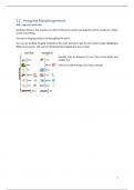
- Spatial resolution
- Temporal resolution
Red circle shows most used methods in developmental biology.
Clinic vs science
Clinical imaging: fast & cheap
In science you go to a really high resolution till even a single molecule.
We focus more on imaging of embryos!
Brief history of imaging
Started with drawings, camera lucida drawing, then in practical course 20 years ago you were given
things to see and had to draw
them, scientists had to be a
bit of an artist. Technology
moved forward to lots of
different microscopies. Big
progress was made with the
discovery of fluorescent dyes
that you could attach to living
things, fluorescent proteins
(GFP). Fluorescence was the
biggest step forward which
permitted us today to watch things live!
- Camera lucida drawing
- Black and white photography-bright
field
- Colour photography-bright field
- Electron microscopy-transmission and
later scanning
- Epi-fluorescence
- Confocal
- Light-sheet
- Cryo-immuno-eEm
- Etc….
- Ultrasound
2
SNR = signal to noise ratio
Doubting Thomas: first scientist, he didn’t believe the stories and legends until he could put a finger
on the actual thing.
Concept of imaging moved a lot throughout the years.
You can use multiple imaging methods in the same embryo to get an even better image highlighting
different structures. Not only for developmental imaging but also in clinic!
typically cells are between 2-5 µm, they can be larger and
smaller tho.
How to see these things, live, living, moving?
1
, 2 very important things:
- Spatial resolution
- Temporal resolution
Red circle shows most used methods in developmental biology.
Clinic vs science
Clinical imaging: fast & cheap
In science you go to a really high resolution till even a single molecule.
We focus more on imaging of embryos!
Brief history of imaging
Started with drawings, camera lucida drawing, then in practical course 20 years ago you were given
things to see and had to draw
them, scientists had to be a
bit of an artist. Technology
moved forward to lots of
different microscopies. Big
progress was made with the
discovery of fluorescent dyes
that you could attach to living
things, fluorescent proteins
(GFP). Fluorescence was the
biggest step forward which
permitted us today to watch things live!
- Camera lucida drawing
- Black and white photography-bright
field
- Colour photography-bright field
- Electron microscopy-transmission and
later scanning
- Epi-fluorescence
- Confocal
- Light-sheet
- Cryo-immuno-eEm
- Etc….
- Ultrasound
2