Cardiac and Smooth Muscle
Cardiac muscle
• Objectives
o Structure of cardiac muscle
o Origin of the heart beat
o Action potential of cardiac muscle
o Regulation of the force of the heart beat
• Muscle
o 3 types of muscle
▪ Skeletal
• Striated – due to regular packing of actin and myosin within the muscle
▪ Smooth
• Irregular
▪ Cardiac
• Striated – due to regular packing of actin and myosin within the muscle
o Muscle fibre
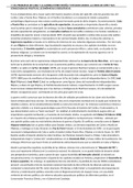
▪ In striated muscle – cardiac and skeletal muscle have a similar structure
▪
o Filament
▪
▪
• Actin-myosin structures = same in skeletal and cardiac muscle
o Binding sites on actin molecules – regulated by calcium binding protein
(troponin)
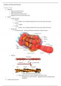
• Cardiac muscle structure
,Cardiac and Smooth Muscle
o
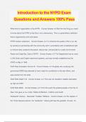
▪ Intercalated discs
• Structural formations between myocardial cells of the heart
• Play roles in bonding cardiac muscles together + transmitting signals between cells
o Attachment points between cardiac muscle cells
• Not present in skeletal muscle
o Skeletal muscle cells fuse together during development = multinucleated
o Cardiac cells are individually nucleated cells → strongly bonded to one
another using intercalated discs
▪ Gap junctions
• Allows cardiac muscle cells to be electrically connected to one another – connect
cytoplasm of cardiac muscle cells
o Cells need to be connected to each other = allowing the heart to beat as a
syncytium – all of the cells beat together
• Ions flow through gap junctions between cells
▪ Attachment proteins
• Desmosomes
o Specialised adhesive proteins that anchor the ends of cardiac muscle fibres
together – so the cells don’t pull apart during contraction
• Contractile activity of a cardiac muscle
o Regular beating of the heart (myogenic activity) – due to an intrinsic pacemaker activity of the heart
o Cardiac muscle
▪ Supplied by nerve fibres that have their origin in the autonomic ganglia
• Autonomic nerves – modulate the rate and force of contraction of cardiac muscle
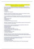
• Cardiac action potential
o
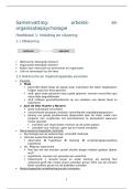
▪ AP in ventricles
• Larger magnitude than skeletal muscle
,Cardiac and Smooth Muscle
• Rapid activation → followed by slow repolarisation phase
▪ AP in atria
• Shorter than AP in ventricles
▪ AP in sinoatrial node
• Sinoatrial node
o Specialised myocardial structure – initiates the electrical impulses to
stimulate contraction
▪ Determines the heart rate
o Found in the superior vena cava and the right atrium
• Slow area of response – slow to reach threshold = to initiate depolarisation (defines
rate at which next action potential will be fired)
o Followed by rapid depolarisation → repolarisation
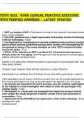
o AP in ventricles
▪
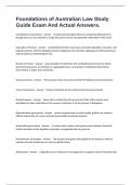
• Action potential – rapid depolarisation → followed by slow repolarisation
• Contraction – action potential – followed by muscle contractions
o Delay between action potential and contraction
• Action potential overlaps the muscle contraction
o Because the action potential is taking a longer period of time – there is
overlap with the contraction = co-incident
▪ No wave summation → no unfused tetani → no tetanus
• During action potential – muscle cannot be reactivated
• Repolarisation time gives a protective time where the
cardiac muscle cannot be activated again
• Shape of response
o Rapid opening of voltage gated Na+ channels – causing depolarisation
o At peak – voltage gated Na+ channels close + voltage-gated Ca2+ channels
open + voltage-gated K+ channels open
▪ Ca2+ + K+ enters cell = repolarisation
• Changing the contraction of cardiac muscle
o Force of contraction of cardiac muscle depends on the degree of stretch of the muscle fibres
▪ More blood entering the heart = the more blood pumped around the body = greater
contraction of cardiac muscle
• Greater stretch of cardiac muscle = greater force of contraction generated by muscle
= more force blood can be pumped around the body
o Regulated by hormonal signals – inotropic response
, Cardiac and Smooth Muscle
▪ Skeletal muscle does not respond to hormonal changes
▪ Cardiac muscle can be regulated by hormones
• Inotropic response
o Responses that will increase the intrinsic contractile component of the heart
o Strength of an individual contraction
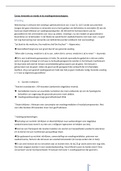
• Length-tension relationship of cardiac muscle
o Stretch muscles = results in greater force of contraction
▪ Active tension – muscle is activated electrically
▪ Resting tension – muscle is stretched
o Muscles have a normal operating range = optimal length of muscles
▪ In cardiac muscle
• Work in normal operating range – below maximal level
o Never under so much stress that they become damaged
▪ In skeletal muscle
• Works in normal operating range – at maximal level
o Can be damaged, but heals over time
• Chronotropic regulation
o Regulation that will alter the rate of the response of a contraction
▪
o Pacemaker activity of the SA node- can be modulated by autonomic nerves
▪ Vagal stimulation
• Vagus = 10th cranial nerve
o Major input from gastrointestinal tract back to
the brain
• After we eat – vagus is stimulated
o Slows heart rate
▪ By reducing the rate at which the SA node reaches threshold
• By opening voltage-gated Na+ channels slower
▪ Sympathetic stimulation
• Sympathetic nervous system – fight or flight response
o Increases heart rate
▪ By increasing the rate at which the SA
node reaches threshold
• By open ing voltage gated Na+ channels quicker
• Excitation-contraction coupling within cardiac muscle – same as skeletal muscle
o Nerve action potential → ACh secretion by nerve ending → end
plate potential → muscle action potential → depolarise T-tubules
and open Ca2+ channels of SR → increase sarcoplasmic Ca2+ →
contraction → pump Ca2+ into SR → relaxation
▪ T-tubules – mostly in ventricular muscle
• Contraction cycle (crossbridge recycling) – same as skeletal muscle
o Myosin heads hydrolyse ATP and become reorientated and
energized
o Myosin heads bind to actin → forming crossbridges
o Myosin crossbridges rotate towards centre of the sarcomere –
power stroke
o As myosin heads bind ATP – crossbridges detach from actin
• Cardiac muscle summary
Cardiac muscle
• Objectives
o Structure of cardiac muscle
o Origin of the heart beat
o Action potential of cardiac muscle
o Regulation of the force of the heart beat
• Muscle
o 3 types of muscle
▪ Skeletal
• Striated – due to regular packing of actin and myosin within the muscle
▪ Smooth
• Irregular
▪ Cardiac
• Striated – due to regular packing of actin and myosin within the muscle
o Muscle fibre
▪ In striated muscle – cardiac and skeletal muscle have a similar structure
▪
o Filament
▪
▪
• Actin-myosin structures = same in skeletal and cardiac muscle
o Binding sites on actin molecules – regulated by calcium binding protein
(troponin)
• Cardiac muscle structure
,Cardiac and Smooth Muscle
o
▪ Intercalated discs
• Structural formations between myocardial cells of the heart
• Play roles in bonding cardiac muscles together + transmitting signals between cells
o Attachment points between cardiac muscle cells
• Not present in skeletal muscle
o Skeletal muscle cells fuse together during development = multinucleated
o Cardiac cells are individually nucleated cells → strongly bonded to one
another using intercalated discs
▪ Gap junctions
• Allows cardiac muscle cells to be electrically connected to one another – connect
cytoplasm of cardiac muscle cells
o Cells need to be connected to each other = allowing the heart to beat as a
syncytium – all of the cells beat together
• Ions flow through gap junctions between cells
▪ Attachment proteins
• Desmosomes
o Specialised adhesive proteins that anchor the ends of cardiac muscle fibres
together – so the cells don’t pull apart during contraction
• Contractile activity of a cardiac muscle
o Regular beating of the heart (myogenic activity) – due to an intrinsic pacemaker activity of the heart
o Cardiac muscle
▪ Supplied by nerve fibres that have their origin in the autonomic ganglia
• Autonomic nerves – modulate the rate and force of contraction of cardiac muscle
• Cardiac action potential
o
▪ AP in ventricles
• Larger magnitude than skeletal muscle
,Cardiac and Smooth Muscle
• Rapid activation → followed by slow repolarisation phase
▪ AP in atria
• Shorter than AP in ventricles
▪ AP in sinoatrial node
• Sinoatrial node
o Specialised myocardial structure – initiates the electrical impulses to
stimulate contraction
▪ Determines the heart rate
o Found in the superior vena cava and the right atrium
• Slow area of response – slow to reach threshold = to initiate depolarisation (defines
rate at which next action potential will be fired)
o Followed by rapid depolarisation → repolarisation
o AP in ventricles
▪
• Action potential – rapid depolarisation → followed by slow repolarisation
• Contraction – action potential – followed by muscle contractions
o Delay between action potential and contraction
• Action potential overlaps the muscle contraction
o Because the action potential is taking a longer period of time – there is
overlap with the contraction = co-incident
▪ No wave summation → no unfused tetani → no tetanus
• During action potential – muscle cannot be reactivated
• Repolarisation time gives a protective time where the
cardiac muscle cannot be activated again
• Shape of response
o Rapid opening of voltage gated Na+ channels – causing depolarisation
o At peak – voltage gated Na+ channels close + voltage-gated Ca2+ channels
open + voltage-gated K+ channels open
▪ Ca2+ + K+ enters cell = repolarisation
• Changing the contraction of cardiac muscle
o Force of contraction of cardiac muscle depends on the degree of stretch of the muscle fibres
▪ More blood entering the heart = the more blood pumped around the body = greater
contraction of cardiac muscle
• Greater stretch of cardiac muscle = greater force of contraction generated by muscle
= more force blood can be pumped around the body
o Regulated by hormonal signals – inotropic response
, Cardiac and Smooth Muscle
▪ Skeletal muscle does not respond to hormonal changes
▪ Cardiac muscle can be regulated by hormones
• Inotropic response
o Responses that will increase the intrinsic contractile component of the heart
o Strength of an individual contraction
• Length-tension relationship of cardiac muscle
o Stretch muscles = results in greater force of contraction
▪ Active tension – muscle is activated electrically
▪ Resting tension – muscle is stretched
o Muscles have a normal operating range = optimal length of muscles
▪ In cardiac muscle
• Work in normal operating range – below maximal level
o Never under so much stress that they become damaged
▪ In skeletal muscle
• Works in normal operating range – at maximal level
o Can be damaged, but heals over time
• Chronotropic regulation
o Regulation that will alter the rate of the response of a contraction
▪
o Pacemaker activity of the SA node- can be modulated by autonomic nerves
▪ Vagal stimulation
• Vagus = 10th cranial nerve
o Major input from gastrointestinal tract back to
the brain
• After we eat – vagus is stimulated
o Slows heart rate
▪ By reducing the rate at which the SA node reaches threshold
• By opening voltage-gated Na+ channels slower
▪ Sympathetic stimulation
• Sympathetic nervous system – fight or flight response
o Increases heart rate
▪ By increasing the rate at which the SA
node reaches threshold
• By open ing voltage gated Na+ channels quicker
• Excitation-contraction coupling within cardiac muscle – same as skeletal muscle
o Nerve action potential → ACh secretion by nerve ending → end
plate potential → muscle action potential → depolarise T-tubules
and open Ca2+ channels of SR → increase sarcoplasmic Ca2+ →
contraction → pump Ca2+ into SR → relaxation
▪ T-tubules – mostly in ventricular muscle
• Contraction cycle (crossbridge recycling) – same as skeletal muscle
o Myosin heads hydrolyse ATP and become reorientated and
energized
o Myosin heads bind to actin → forming crossbridges
o Myosin crossbridges rotate towards centre of the sarcomere –
power stroke
o As myosin heads bind ATP – crossbridges detach from actin
• Cardiac muscle summary