Unit 21 LA A&B Radiation use in medical diagnosis and treatment Unknown
Radiation use in medical diagnosis and treatment
Intro
I have secured a placement within the Radiography Department of a teaching hospital.
I'll be able to learn a lot about how ionising and non-ionizing radiation are used to
diagnose and treat a range of disorders during my clinical placement by observing the
work of a certified radiographer. I will write a report at the end of my placement to
demonstrate my knowledge in university interviews.
✔
Non-ionising radiation
Any electromagnetic radiation that does not have enough energy to ionise atoms or
molecules, making electrons oscillate instead of being removed and raising body
temperature is referred to as non-ionizing radiation (Díez). They are waves with a long
wavelength, low frequency, and low energy(Díez).
Waves can be produced and detected by many technologies. In an MRI, waves are
absorbed by atoms and subatomic particles, which then release radiation. Various
substances reflect ultrasonic waves.Light waves that are focused produce energy.
Infrared thermography
Infrared thermography is an example of a medical device using non-ionising radiation. A
thermal imager is used to detect radiation (heat) flowing from an object, convert it to
temperature, and display an image of the temperature distribution (Trout). This process
is known as infrared thermography (Trout). Thermograms illustrations that detect
temperature distribution allow the observation of heat-producing objects that are
invisible to the eyepiece (Trout). It is frequently used in condition monitoring and
predictive maintenance (Trout).The wavelength of infrared radiation, a form of non-
ionizing radiation, can range from 700 nm to 1000000nms.
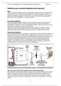
(“Schematic diagram of infrared thermography camera. | Download Scientific Diagram”)
Figure 1 shows an infrared thermography camera's labelled operation:
This image shows the process it takes to end up with a real time display as you can see
from the image, first the object emits IR energy and that energy is collected and then
filtered so it passes through a selected spectral band. After this the detector then
converts IR energy to an electric signal then the amplifier and the signal processing
converts the electric signal to a thermography. Which is then displayed on a real time
display.
1
,Unit 21 LA A&B Radiation use in medical diagnosis and treatment Unknown
Part A.P1
(“Infrared Lab Members [IMAGE]”)
Figure 2: An example of an infrared photo
An area with more infrared waves appears more yellow or red in infrared images, while
an area with less infrared waves appears blue or purple.
There are multiple medical uses for infrared thermography. One way they can be used is
to diagnose cancers, for example breast cancer (“Medical applications of infrared
thermography: A review”).This is a result of breast cancer heating up the skin, which the
infrared thermography camera can identify. This would result in a temperature shift
because if a tumour were to develop in the body, it would construct its own blood
vessels. Cell growth to make the blood vessels would produce heat, which the infrared
thermography camera would be able to detect. Fever detection is another medical use
for infrared thermography. This is carried out because a patient's body temperature
change mostly causes the early stages of a fever.An infrared thermometer can detect
this change in temperature because if the body temperature is high, more infrared
waves are emitted and detected by the thermometer.Due to their precision in detecting
heat, infrared thermometers are used to detect fevers. Due to the risk of mercury
spilling out if a mercury thermometer breaks since mercury is corrosive, infrared
cameras are also used in thermometers.
There are many advantages of infrared thermography. One advantage in infrared
thermography is that its non-contact (“Infrared cameras for use in medicine |
InfraTec”). This is important because the detector doesn't touch the skin. Another
advantage is that this device does not produce heat; it just detects heat.This is beneficial
since there would be a risk of burns if heat were produced during the treatment.Another
benefit of infrared thermography is its high precision. This is helpful because it provides
for a higher level of diagnostic accuracy by accurately detecting the degree of heat. One
limitation of infrared thermography is that to interpret the results you have to be
required a certain amount of experience and knowledge (“Infrared Thermography and
Energy Efficiency”). Another limitation is that it's expensive. This is a limitation as the
cost might prevent some individuals from buying it.
Part A.P2
MRI
Magnetic resonance imaging scans are yet another medical technology that uses non-
ionizing radiation (MRI). A specific kind of scan that creates highly detailed images of the
2
, Unit 21 LA A&B Radiation use in medical diagnosis and treatment Unknown
interior of the body using radio waves and powerful magnetic fields (“Overview - - - MRI
scan”).The electromagnetic spectrum contains non-ionizing radiation, including radio
waves. In the electromagnetic spectrum, radio waves are the least energetic and have
the longest wavelengths.Radio waves' long wavelengths makes them suitable for
communication, and this feature can be applied to the field of medicine.
Main MRI Head coil
superconducti Scanner
ng
coil
Gradient
and Active Patient
Shim coils
Patient
Radio table
frequency
(body) cod
Field
Figure 3: what an MRI scan looks like
(Jenkinson and Chappell)
The patient's body is put in a strong magnetic field to start the MRI scan.The hydrogen
atoms in the body start to resonate, as shown in figure 4. The protons in the hydrogen
atoms begin to spin more as a result, which changes their axis and aligns it with the
magnetic field. Now they take up some of the energy released.The signal is then turned
off, and the protons resume their resting state. This causes them to generate energy
(radio waves), which can be used to create an image of the body's inside.
Protons in the Protons in the
body MRI scanner
Hydrogen
proton
No magnetic 80 magnetic
H20 field field
Molecules
Rf
pulse
Figure 4: stages in an MRI scan (Britt et al.)Part A.P1
3
Radiation use in medical diagnosis and treatment
Intro
I have secured a placement within the Radiography Department of a teaching hospital.
I'll be able to learn a lot about how ionising and non-ionizing radiation are used to
diagnose and treat a range of disorders during my clinical placement by observing the
work of a certified radiographer. I will write a report at the end of my placement to
demonstrate my knowledge in university interviews.
✔
Non-ionising radiation
Any electromagnetic radiation that does not have enough energy to ionise atoms or
molecules, making electrons oscillate instead of being removed and raising body
temperature is referred to as non-ionizing radiation (Díez). They are waves with a long
wavelength, low frequency, and low energy(Díez).
Waves can be produced and detected by many technologies. In an MRI, waves are
absorbed by atoms and subatomic particles, which then release radiation. Various
substances reflect ultrasonic waves.Light waves that are focused produce energy.
Infrared thermography
Infrared thermography is an example of a medical device using non-ionising radiation. A
thermal imager is used to detect radiation (heat) flowing from an object, convert it to
temperature, and display an image of the temperature distribution (Trout). This process
is known as infrared thermography (Trout). Thermograms illustrations that detect
temperature distribution allow the observation of heat-producing objects that are
invisible to the eyepiece (Trout). It is frequently used in condition monitoring and
predictive maintenance (Trout).The wavelength of infrared radiation, a form of non-
ionizing radiation, can range from 700 nm to 1000000nms.
(“Schematic diagram of infrared thermography camera. | Download Scientific Diagram”)
Figure 1 shows an infrared thermography camera's labelled operation:
This image shows the process it takes to end up with a real time display as you can see
from the image, first the object emits IR energy and that energy is collected and then
filtered so it passes through a selected spectral band. After this the detector then
converts IR energy to an electric signal then the amplifier and the signal processing
converts the electric signal to a thermography. Which is then displayed on a real time
display.
1
,Unit 21 LA A&B Radiation use in medical diagnosis and treatment Unknown
Part A.P1
(“Infrared Lab Members [IMAGE]”)
Figure 2: An example of an infrared photo
An area with more infrared waves appears more yellow or red in infrared images, while
an area with less infrared waves appears blue or purple.
There are multiple medical uses for infrared thermography. One way they can be used is
to diagnose cancers, for example breast cancer (“Medical applications of infrared
thermography: A review”).This is a result of breast cancer heating up the skin, which the
infrared thermography camera can identify. This would result in a temperature shift
because if a tumour were to develop in the body, it would construct its own blood
vessels. Cell growth to make the blood vessels would produce heat, which the infrared
thermography camera would be able to detect. Fever detection is another medical use
for infrared thermography. This is carried out because a patient's body temperature
change mostly causes the early stages of a fever.An infrared thermometer can detect
this change in temperature because if the body temperature is high, more infrared
waves are emitted and detected by the thermometer.Due to their precision in detecting
heat, infrared thermometers are used to detect fevers. Due to the risk of mercury
spilling out if a mercury thermometer breaks since mercury is corrosive, infrared
cameras are also used in thermometers.
There are many advantages of infrared thermography. One advantage in infrared
thermography is that its non-contact (“Infrared cameras for use in medicine |
InfraTec”). This is important because the detector doesn't touch the skin. Another
advantage is that this device does not produce heat; it just detects heat.This is beneficial
since there would be a risk of burns if heat were produced during the treatment.Another
benefit of infrared thermography is its high precision. This is helpful because it provides
for a higher level of diagnostic accuracy by accurately detecting the degree of heat. One
limitation of infrared thermography is that to interpret the results you have to be
required a certain amount of experience and knowledge (“Infrared Thermography and
Energy Efficiency”). Another limitation is that it's expensive. This is a limitation as the
cost might prevent some individuals from buying it.
Part A.P2
MRI
Magnetic resonance imaging scans are yet another medical technology that uses non-
ionizing radiation (MRI). A specific kind of scan that creates highly detailed images of the
2
, Unit 21 LA A&B Radiation use in medical diagnosis and treatment Unknown
interior of the body using radio waves and powerful magnetic fields (“Overview - - - MRI
scan”).The electromagnetic spectrum contains non-ionizing radiation, including radio
waves. In the electromagnetic spectrum, radio waves are the least energetic and have
the longest wavelengths.Radio waves' long wavelengths makes them suitable for
communication, and this feature can be applied to the field of medicine.
Main MRI Head coil
superconducti Scanner
ng
coil
Gradient
and Active Patient
Shim coils
Patient
Radio table
frequency
(body) cod
Field
Figure 3: what an MRI scan looks like
(Jenkinson and Chappell)
The patient's body is put in a strong magnetic field to start the MRI scan.The hydrogen
atoms in the body start to resonate, as shown in figure 4. The protons in the hydrogen
atoms begin to spin more as a result, which changes their axis and aligns it with the
magnetic field. Now they take up some of the energy released.The signal is then turned
off, and the protons resume their resting state. This causes them to generate energy
(radio waves), which can be used to create an image of the body's inside.
Protons in the Protons in the
body MRI scanner
Hydrogen
proton
No magnetic 80 magnetic
H20 field field
Molecules
Rf
pulse
Figure 4: stages in an MRI scan (Britt et al.)Part A.P1
3