BACTERIAL PATHOGENESIS AND INFECTIONS
I. Adhesion and colonisation: Attachment (weak, reversible) → Adhesion (specific, irreversible, receptor-adhesin) → aggregation → Dispersion
- Attachment: mediated by ionic interaction (hydrogen bonding & Waal’s force), cell surfaces have a net negative charge.
- Adhesion: causes loss of microvilli from enterocytes and changes in actin cytoskeleton signalling pathways following receptor’s phosphorylation. Example: uropathogenic E.
coli: P Fimbriae + Gal(α 1-4) Gal; N.meningitidis Type IV fimbriae + heparin sulphate or Opa proteins + integrins; S. aureus: MSCRAMMs + ECM proteins (fibronectin, fibrinogen)
- Aggregation: biofilm or conditioning films. Example: PIA of Sa is encoded on ica locus, contains eDNA, is a beta N-acetyl glucosamine
Invasion: adhesion (triggers host signal transduction pathways) → invasins → b-integrins → integrins mediate attachment of cells to ECM. Example of integrin: cell surface
receptor (on Legionella pneumophila) for complement component C3bi on macrophage surface.
Successful colonization involves: Adhesion; Nutrient acquisition; Avoidance of host defences (capsule & IgA protease production); Replication
II. Protein secretion
Sec: post-translation secretion (SecA,B)/ co-translational (SRP)
T7SS: Mycobacteria
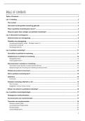
- T3SS: needle
complex, mediates
invasion, inhibits
endosome
maturation &
phagocytosis by
macrophages,
intimate adhesion
(EPEC/EHEC).
Example:
III. Toxins: specific poison, albuminous nature, acts in pore formation, binding, phospholipase, ADP ribosylation, monoglycosylation, proteolysis
1) Diphtheria: caused by C. diphtheriae, mediates ADP-ribosylation of EF-2 → proteins cannot be made → kill the cell.
Tox expression regulated by iron-dependent repressor (DtxR)/ When iron conc. Is low, DtxR detaches from the operator and tox is expressed.
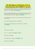
2) Cholera toxin: binds to plasma membrane of intestinal epithelial cells → causes ADP-ribosylation of Gs protein (a stimulator of adenylate cyclase) → constantly produces c-AMP
from ATP → c-AMP mediates cellular responses, e.g., Cl- efflux in the intestinal lumen.
3) Pertussis: caused by Bordetella pertussis which colonise ciliated cells of respiratory mucosa; toxin has receptor-binding & catalytic activity.
Targets Gi protein (inhibitor of adenylate cyclase) → over-production of cAMP affects normal biological signals → neurological disorders causing cough → dissemination of
pathogens in the environment.
4) Botulinum: by C. botulinum, targets the neurons between neuro-muscular junctions, cleaving SNARE proteins → synaptic vesicles can’t fuse w/t membrane → neurotransmitters
can’t be released & tell the muscle to contract → paralysis. >< tetanus targets inhibitory neurons, by C. tetani.
, 5) Endotoxins: LOS (lacks O-Antigen) by N. meningitidis infects through nasopharynx and invades bloodstream → meningococcemia and meningitis.
IV. Iron and Infections: Iron is essential nutrients for bacterial growth but free iron is toxic, causing damage for DNA and cell membranes.
- Iron limitation in the body is controlled by: hepcidin secreted when extracellular iron level is high (internalize iron exporter ferroportin, reducing free iron); hypoferraemia:
reduce iron uptake in the intestine, increase ferritin synthesis in the liver, release apo-lactoferrin by neutrophils, suppress iron efflux from macrophages, release haemopexin and
haptoglobin in the liver; haemochromatosis: increased iron uptake in genetic disorders e.g. thalassaemia, leukaemia, anaemia.
- Bacteria whose virulence enhanced by iron: K. pneumoniae, N. gonorrhoeae, Pa, Sa, V.cholerae, Clostridium perfringens, E.coli, L. monocytogenes.
- Iron acquisition: cleavage of iron-binding site; Fe3+ to Fe2+; production & export of Iron Chelators (Siderophores); expression of surface receptor.
E. coli expresses a receptor for a fungal siderophore called ferrichrome. Several OTM receptors for ferri-siderophores are targets for colicin antibiotics.
- Haem as iron sources: IsdA, hts (Sa); Shp,HtsA (S.pyogenes); PiuA (S.pneumonia); HupC (L.monocytogenes)
- Haemophores: are groups of secreted haem-binding proteins; a specific receptor on bacterial surfaces (e.g. erratia marcescens, Pseudomonas aeruginosa, Yersinia pestis,
Haemophilus influenzae, Bacillus anthracis)
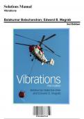
V. Biofilm: Non-motile = increased biofilm = more chronic infection; promoted by cyclic-di-GMP. Step 1: Aggregation & attachment involves adhesins mediate cell-cell, -
surface or -polymer interactions. Step 2: Growth & accumulation: by replication, synthesis of EPS/eDNA & others → more antimicrobial tolerant, QS activated. Step 3:
Disaggregation, detachment, & dispersal: due to mechanical env stress, exploration of other niches, chemical treatment (NO → upregulates PPD).
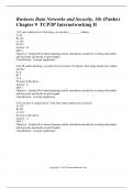
Gac/Rsmsystem: GacS -> GacA-(P) → transcribed RsmZ & RsmY → bind to RsmA → RsmA dissociation from
RBS activates pel, psl, hcnABC, phz, T6SS
Cyclic-di-GMP: synthesized by di-guanylate cyclase and degraded by Phosphodiesterase
Antimicrobial tolerance: reduced growth, decreased uptake of antibiotics, depletion of antibiotic
targets; efflux pump expression, b-lactamase expression, impaired penetration.
Inhibition of opsonization, phagocytosis, complement deposition, AMPs by EPS & eDNA
VI. Quorum sensing : (+) bacterial Autoinducing peptides: lantibiotics, linear/cyclic peptides e.g.,
ComC, thiolactones (Staphylococci), lactones (Enterococci).
QS inhibitor targets: triclosan inhibits fatty acid synthesis & C4-HSL synthesis, lactonases &
acyclases/amidases cleave AHL signal molecules, N- acylhomoserine lactone degrading enzymes, PqsR
inhibitors, peptide nucleic acids (PNAs) block translation of pqsA.
QS inhibitor’s disadvantages: narrow spectrum, non-bactericidal, diagnostic system
I. Adhesion and colonisation: Attachment (weak, reversible) → Adhesion (specific, irreversible, receptor-adhesin) → aggregation → Dispersion
- Attachment: mediated by ionic interaction (hydrogen bonding & Waal’s force), cell surfaces have a net negative charge.
- Adhesion: causes loss of microvilli from enterocytes and changes in actin cytoskeleton signalling pathways following receptor’s phosphorylation. Example: uropathogenic E.
coli: P Fimbriae + Gal(α 1-4) Gal; N.meningitidis Type IV fimbriae + heparin sulphate or Opa proteins + integrins; S. aureus: MSCRAMMs + ECM proteins (fibronectin, fibrinogen)
- Aggregation: biofilm or conditioning films. Example: PIA of Sa is encoded on ica locus, contains eDNA, is a beta N-acetyl glucosamine
Invasion: adhesion (triggers host signal transduction pathways) → invasins → b-integrins → integrins mediate attachment of cells to ECM. Example of integrin: cell surface
receptor (on Legionella pneumophila) for complement component C3bi on macrophage surface.
Successful colonization involves: Adhesion; Nutrient acquisition; Avoidance of host defences (capsule & IgA protease production); Replication
II. Protein secretion
Sec: post-translation secretion (SecA,B)/ co-translational (SRP)
T7SS: Mycobacteria
- T3SS: needle
complex, mediates
invasion, inhibits
endosome
maturation &
phagocytosis by
macrophages,
intimate adhesion
(EPEC/EHEC).
Example:
III. Toxins: specific poison, albuminous nature, acts in pore formation, binding, phospholipase, ADP ribosylation, monoglycosylation, proteolysis
1) Diphtheria: caused by C. diphtheriae, mediates ADP-ribosylation of EF-2 → proteins cannot be made → kill the cell.
Tox expression regulated by iron-dependent repressor (DtxR)/ When iron conc. Is low, DtxR detaches from the operator and tox is expressed.
2) Cholera toxin: binds to plasma membrane of intestinal epithelial cells → causes ADP-ribosylation of Gs protein (a stimulator of adenylate cyclase) → constantly produces c-AMP
from ATP → c-AMP mediates cellular responses, e.g., Cl- efflux in the intestinal lumen.
3) Pertussis: caused by Bordetella pertussis which colonise ciliated cells of respiratory mucosa; toxin has receptor-binding & catalytic activity.
Targets Gi protein (inhibitor of adenylate cyclase) → over-production of cAMP affects normal biological signals → neurological disorders causing cough → dissemination of
pathogens in the environment.
4) Botulinum: by C. botulinum, targets the neurons between neuro-muscular junctions, cleaving SNARE proteins → synaptic vesicles can’t fuse w/t membrane → neurotransmitters
can’t be released & tell the muscle to contract → paralysis. >< tetanus targets inhibitory neurons, by C. tetani.
, 5) Endotoxins: LOS (lacks O-Antigen) by N. meningitidis infects through nasopharynx and invades bloodstream → meningococcemia and meningitis.
IV. Iron and Infections: Iron is essential nutrients for bacterial growth but free iron is toxic, causing damage for DNA and cell membranes.
- Iron limitation in the body is controlled by: hepcidin secreted when extracellular iron level is high (internalize iron exporter ferroportin, reducing free iron); hypoferraemia:
reduce iron uptake in the intestine, increase ferritin synthesis in the liver, release apo-lactoferrin by neutrophils, suppress iron efflux from macrophages, release haemopexin and
haptoglobin in the liver; haemochromatosis: increased iron uptake in genetic disorders e.g. thalassaemia, leukaemia, anaemia.
- Bacteria whose virulence enhanced by iron: K. pneumoniae, N. gonorrhoeae, Pa, Sa, V.cholerae, Clostridium perfringens, E.coli, L. monocytogenes.
- Iron acquisition: cleavage of iron-binding site; Fe3+ to Fe2+; production & export of Iron Chelators (Siderophores); expression of surface receptor.
E. coli expresses a receptor for a fungal siderophore called ferrichrome. Several OTM receptors for ferri-siderophores are targets for colicin antibiotics.
- Haem as iron sources: IsdA, hts (Sa); Shp,HtsA (S.pyogenes); PiuA (S.pneumonia); HupC (L.monocytogenes)
- Haemophores: are groups of secreted haem-binding proteins; a specific receptor on bacterial surfaces (e.g. erratia marcescens, Pseudomonas aeruginosa, Yersinia pestis,
Haemophilus influenzae, Bacillus anthracis)
V. Biofilm: Non-motile = increased biofilm = more chronic infection; promoted by cyclic-di-GMP. Step 1: Aggregation & attachment involves adhesins mediate cell-cell, -
surface or -polymer interactions. Step 2: Growth & accumulation: by replication, synthesis of EPS/eDNA & others → more antimicrobial tolerant, QS activated. Step 3:
Disaggregation, detachment, & dispersal: due to mechanical env stress, exploration of other niches, chemical treatment (NO → upregulates PPD).
Gac/Rsmsystem: GacS -> GacA-(P) → transcribed RsmZ & RsmY → bind to RsmA → RsmA dissociation from
RBS activates pel, psl, hcnABC, phz, T6SS
Cyclic-di-GMP: synthesized by di-guanylate cyclase and degraded by Phosphodiesterase
Antimicrobial tolerance: reduced growth, decreased uptake of antibiotics, depletion of antibiotic
targets; efflux pump expression, b-lactamase expression, impaired penetration.
Inhibition of opsonization, phagocytosis, complement deposition, AMPs by EPS & eDNA
VI. Quorum sensing : (+) bacterial Autoinducing peptides: lantibiotics, linear/cyclic peptides e.g.,
ComC, thiolactones (Staphylococci), lactones (Enterococci).
QS inhibitor targets: triclosan inhibits fatty acid synthesis & C4-HSL synthesis, lactonases &
acyclases/amidases cleave AHL signal molecules, N- acylhomoserine lactone degrading enzymes, PqsR
inhibitors, peptide nucleic acids (PNAs) block translation of pqsA.
QS inhibitor’s disadvantages: narrow spectrum, non-bactericidal, diagnostic system