Emilia Hawkins
Unit 8: Physiology of Human Body Systems
C: Explore the physiology of the digestive system and the use of corrective treatments for dietary
related diseases.
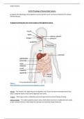
A diagram showing the role of each organs of the digestive system.
Oesophagus
https://
www.britannica.com/science/human-digestive-system
Mouth – The mouth is the beginning of the digestive tract, food is chewed and broken down into
pieces using the teeth so that can be digested more easily.
Tongue – The tongue assists swallowing and encourages food movement during chewing.
Salivary glands – The salivary glands produce saliva which allows food to be swallowed more easily.
It also contains enzymes which helps the stomach to break down large molecules.
, Emilia Hawkins
Pharynx – The pharynx carries food and fluids down from the mouth by using an automatic
swallowing reflex that is caused by sensory receptors. The epiglottis directs the food into the
oesophagus and prevents it from going into the larynx and trachea.
Oesophagus – The oesophagus connects the throat with the stomach and transports food by
peristalsis by alternating contraction and relaxation of the muscles. It is lined with pink tissue called
mucosa and has glands which secrete mucus to allow food to pass through more easily.
Stomach – The stomach receives food from the oesophagus and aids in digestion by secreting a
strong acid and enzymes which break down the food. The muscles in the walls of the stomach
contract periodically to enhance digestion.
Small intestines – Food passes from the stomach and into the small intestine using the pyloric
sphincter. It is made up of 3 sections called the duodenum, jejunum and ileum. The duodenum
mixes food from the stomach with enzymes that come from the pancreas and bile, which is stored in
the gallbladder, which further helps in breaking down the food. The walls of the jejunum absorb
nutrients using a large surface area due to the many circular folds. The ileum absorbs bile and
vitamin B12.
Pancreas – The pancreas releases digestive enzymes into the last part of the small intestine to help
break down carbohydrates, fats and proteins. It also produced insulin which controls the blood sugar
levels and metabolism.
Liver – The liver produces bile and purifies blood that contains nutrients which have been absorbed
from the small intestine.
Gall bladder – The gallbladder concentrates and stores bile and also releases it into the small
intestine, which neutralises stomach acid and aids in the absorption and digestion of fats.
Large intestines – The large intestine is responsible for processing waste and is made up of the
cecum, the ascending colon, the transverse colon, the descending colon and the sigmoid colon. It
does not contain villi or cells that secrete digestive enzymes but does contain goblet cells that
secrete mucus to protect the colon from acids and gases.
Rectum– The rectum receives faeces from the colon and holds it there until it can be evacuated. The
sensors in the wall of the rectum send signals to the brain and the can brain makes the voluntary
decision to relax the sphincters in the anus.
Anus – The anus is the last part of the digestive system and it contains two sphincter muscles
(internal and external) and pelvic floor muscles which control the movement of faeces.
https://my.clevelandclinic.org/health/body/7041-digestive-system
Practical analytical tests to establish the nutritional content of a variety of foods.
The Iodine test for starch
Method:
1. Put some of the food sample into a test tube.
2. Add a few drops of iodine solution to the food sample using a pipette.
3. If starch is present, the solution will turn from brown to blue-black.
Unit 8: Physiology of Human Body Systems
C: Explore the physiology of the digestive system and the use of corrective treatments for dietary
related diseases.
A diagram showing the role of each organs of the digestive system.
Oesophagus
https://
www.britannica.com/science/human-digestive-system
Mouth – The mouth is the beginning of the digestive tract, food is chewed and broken down into
pieces using the teeth so that can be digested more easily.
Tongue – The tongue assists swallowing and encourages food movement during chewing.
Salivary glands – The salivary glands produce saliva which allows food to be swallowed more easily.
It also contains enzymes which helps the stomach to break down large molecules.
, Emilia Hawkins
Pharynx – The pharynx carries food and fluids down from the mouth by using an automatic
swallowing reflex that is caused by sensory receptors. The epiglottis directs the food into the
oesophagus and prevents it from going into the larynx and trachea.
Oesophagus – The oesophagus connects the throat with the stomach and transports food by
peristalsis by alternating contraction and relaxation of the muscles. It is lined with pink tissue called
mucosa and has glands which secrete mucus to allow food to pass through more easily.
Stomach – The stomach receives food from the oesophagus and aids in digestion by secreting a
strong acid and enzymes which break down the food. The muscles in the walls of the stomach
contract periodically to enhance digestion.
Small intestines – Food passes from the stomach and into the small intestine using the pyloric
sphincter. It is made up of 3 sections called the duodenum, jejunum and ileum. The duodenum
mixes food from the stomach with enzymes that come from the pancreas and bile, which is stored in
the gallbladder, which further helps in breaking down the food. The walls of the jejunum absorb
nutrients using a large surface area due to the many circular folds. The ileum absorbs bile and
vitamin B12.
Pancreas – The pancreas releases digestive enzymes into the last part of the small intestine to help
break down carbohydrates, fats and proteins. It also produced insulin which controls the blood sugar
levels and metabolism.
Liver – The liver produces bile and purifies blood that contains nutrients which have been absorbed
from the small intestine.
Gall bladder – The gallbladder concentrates and stores bile and also releases it into the small
intestine, which neutralises stomach acid and aids in the absorption and digestion of fats.
Large intestines – The large intestine is responsible for processing waste and is made up of the
cecum, the ascending colon, the transverse colon, the descending colon and the sigmoid colon. It
does not contain villi or cells that secrete digestive enzymes but does contain goblet cells that
secrete mucus to protect the colon from acids and gases.
Rectum– The rectum receives faeces from the colon and holds it there until it can be evacuated. The
sensors in the wall of the rectum send signals to the brain and the can brain makes the voluntary
decision to relax the sphincters in the anus.
Anus – The anus is the last part of the digestive system and it contains two sphincter muscles
(internal and external) and pelvic floor muscles which control the movement of faeces.
https://my.clevelandclinic.org/health/body/7041-digestive-system
Practical analytical tests to establish the nutritional content of a variety of foods.
The Iodine test for starch
Method:
1. Put some of the food sample into a test tube.
2. Add a few drops of iodine solution to the food sample using a pipette.
3. If starch is present, the solution will turn from brown to blue-black.