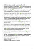
Blood
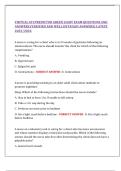
Four components:
- Plasma water 92%, sugar, fat, proteins and salts. Transport
- Red blood cells (erythrocytes) 40 – 45% no nucleus, hemoglobin. Haematocrit number
of red blood cells.
- White blood cells (leukocytes) 1 % protect
o Monocytes 3 – 8 % can develop into macrophages
o Lymphocytes 25%
T-lymphocytes regulate other immune cells and directly attack
B-lymphocytes makes antibodies.
o Granulocytes contain cytoplasmic inclusions
Neutrophils 55 – 70 % body bacteria slayers, number increase explosively.
Eosinophils 2 – 4 % lead counterattack against multicellular parasites.
Basophils 0,5 – 1 % dilated and attracts other leukocytes with histamine.
Causes vasodilation. Secrete chemicals to help with immune system.
- Platelets (thrombocytes)
blood coagulation. Fibrin clot
made, which covers wound.
MOETEN HERKENNEN!
Three functions:
- Distribution oxygen/dioxide, metabolic waste, hormones.
- Regulation body temperature, pH, adequate fluid volume.
- Protection preventing blood loss and infections.
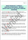
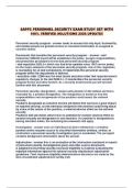
Origin of blood cells (haematopoiesis)
Blood cells are produced in the bone marrow.
Haematopoiesis is controlled by cytokines, which are
peptides or proteins released from one cell that affect
the growth or activity of another cell.
EPO (erythropoietin) produced in kidney, controls
red blood cell synthesis. Stimulus for synthesis is low
oxygen levels, which stimulate HIF-1, which turns on
the EPO gene.
TPO (thrombopoietin) produced in liver , controls
megakaryocytes (become platelets)
Colony-stimulating factors, stem cell factors, interleukins produced by endothelium and fibroblast
of bone marrow, and leukocytes. Regulate all types of blood cells.
IL-3 (interleukin) controls white blood cell synthesis.
Plasma and interstitial fluid
Plasma contains higher concentration of oxygen and proteins. Present in blood.
Interstitial fluid contains higher concentration of carbon dioxide. Present between tissue and
capillaries.
,Transport
Transcellular through cell.
Paracellular across tight junctions.
Passive (diffusion) down concentration gradient. Rate of transport determined by:
- Electrochemical gradient for diffusion across cell membrane.
- Permeability of membrane.
- Time that fluid containing substance remains within tubules.
Simple diffusion nonpolar and lipid-soluble substances diffuse through lipid bilayer
Carrier mediated facilitated diffusion bind to protein carriers in membrane or through water filled
protein channels.
Osmosis diffusion of solution through selective permeable membrane.
Active against electrochemical gradient which requires
energy.
Primary active transport directly coupled to energy provided by
ATP. Using ATPase’s.
Secondary active transport indirectly coupled to energy. Two or
more substances cross membrane via transporter together. Energy
released is used for other substance against electrochemical
gradient. Often involves counter-transport.
Pinocytosis molecules absorbed via vesicles.
Antiporter co-transporter and membrane protein in secondary
transport that transport two or more different molecules across
membrane.
Uniporter transport protein that can only transport one substrate.
Symporter transport protein transport two substances same direction.
Transcytosis endocytosis and exocytosis in same cell.
Co-transport diffusion energy of one substance that can pull other substances along with them
through the cell membrane.
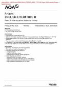
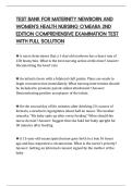
,Autonomic system
Parasympathetic nervous system Sympathetic nervous system
Nerves arise from the bulbus to different Come out of spinal cord the synapse in
organs. Nerve X goes to heart, lungs, and other ganglia.
organs. This nerve is called the vagal nerve. - Short pre-ganglionic nerves (to synapse)
- Long pre-parasymptonic nerves - Long post-ganglionic nerves (to organ)
- Short post-parasymptonic nerves
Rest – digest Anabolic Action – fight-or-flight response Catabolic
Acetylcholine (Ach) as transmitter in post- Noradrenaline (NA) as transmitter in post-
ganglionic neurons. ganglionic neurons.
All 1st connections use Ach as neurotransmitter.
Acetylcholine different receptors are involved
Nicotine (ion channels)
- Nm neuro muscular ending
- Nn autonomic ganglia adrenal gland
Muscarine (G-protein coupled receptors, GPCRs) activated by Ach.
- M1 autonomic ganglia EPSP
- M2 heart presynaptic
- M3 smooth muscle + glands
Adrenergic nervous system
- Noradrenaline (misses one methyl group)
o Alpha-receptors
α1-receptor blood vessel
α2-receptor presynaptic nerve ending
- Adrenaline
o Beta-receptors
β1-receptor heart, kidney
β2-receptor lungs, arteries, uterus, neuro-musc. Ending
salbutamol short acting β2-anagonist (broken down rapidly)
, salmeterol long acting β2-anagonist (broken down slowly)
β3-receptor fat cells.
Adrenal medulla nerve cells located in medulla makes adrenaline.
Phenylethanolamine-N-methyltransferase converts NA into A (adding methyl-group) in cytoplasm.
Varicosities around blood vessels can release NA NA to α1-receptor (contraction) or β2-receptor
(dilation).
- No true synaptic cleft
o NA extravascular side (outside of the artery or vein)
o A intravascular side (inside of artery or vein)
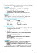
Cholinergic synapse
Choline + AcCoA Ach + CoA
- Stored in vesicles fuse in membrane when Ca++ is high (presynaptic membrane)
Ach is very rapidly broken down by AchE, the choline is taken up for reuse.
Pre-synaptic control (negative feedback)
- Ach couples to M2 ATP cAMP less calcium.
- NA can activate α2-receptors also inhibits calcium.
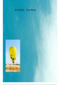
Adrenergic synapse
Tyrosine DOPA Dopamine NA
Ca++ will result in the exocytosis of NA Stimulates α- and β-receptors
NA 10% taken up and broken down by COMT (catechol-O-methyltransferase)
NA 90% taken up by NA-transporters and pumped into the cell
- NA transport into vesicle to refill
- Broken down by MAO (monoamine-oxidase) into inactive substances.
Pre-synaptic control
- α-receptors and M2-receptors give negative feedback control produce Ca ++
- α2 or β2 have positive effect
Four components:
- Plasma water 92%, sugar, fat, proteins and salts. Transport
- Red blood cells (erythrocytes) 40 – 45% no nucleus, hemoglobin. Haematocrit number
of red blood cells.
- White blood cells (leukocytes) 1 % protect
o Monocytes 3 – 8 % can develop into macrophages
o Lymphocytes 25%
T-lymphocytes regulate other immune cells and directly attack
B-lymphocytes makes antibodies.
o Granulocytes contain cytoplasmic inclusions
Neutrophils 55 – 70 % body bacteria slayers, number increase explosively.
Eosinophils 2 – 4 % lead counterattack against multicellular parasites.
Basophils 0,5 – 1 % dilated and attracts other leukocytes with histamine.
Causes vasodilation. Secrete chemicals to help with immune system.
- Platelets (thrombocytes)
blood coagulation. Fibrin clot
made, which covers wound.
MOETEN HERKENNEN!
Three functions:
- Distribution oxygen/dioxide, metabolic waste, hormones.
- Regulation body temperature, pH, adequate fluid volume.
- Protection preventing blood loss and infections.
Origin of blood cells (haematopoiesis)
Blood cells are produced in the bone marrow.
Haematopoiesis is controlled by cytokines, which are
peptides or proteins released from one cell that affect
the growth or activity of another cell.
EPO (erythropoietin) produced in kidney, controls
red blood cell synthesis. Stimulus for synthesis is low
oxygen levels, which stimulate HIF-1, which turns on
the EPO gene.
TPO (thrombopoietin) produced in liver , controls
megakaryocytes (become platelets)
Colony-stimulating factors, stem cell factors, interleukins produced by endothelium and fibroblast
of bone marrow, and leukocytes. Regulate all types of blood cells.
IL-3 (interleukin) controls white blood cell synthesis.
Plasma and interstitial fluid
Plasma contains higher concentration of oxygen and proteins. Present in blood.
Interstitial fluid contains higher concentration of carbon dioxide. Present between tissue and
capillaries.
,Transport
Transcellular through cell.
Paracellular across tight junctions.
Passive (diffusion) down concentration gradient. Rate of transport determined by:
- Electrochemical gradient for diffusion across cell membrane.
- Permeability of membrane.
- Time that fluid containing substance remains within tubules.
Simple diffusion nonpolar and lipid-soluble substances diffuse through lipid bilayer
Carrier mediated facilitated diffusion bind to protein carriers in membrane or through water filled
protein channels.
Osmosis diffusion of solution through selective permeable membrane.
Active against electrochemical gradient which requires
energy.
Primary active transport directly coupled to energy provided by
ATP. Using ATPase’s.
Secondary active transport indirectly coupled to energy. Two or
more substances cross membrane via transporter together. Energy
released is used for other substance against electrochemical
gradient. Often involves counter-transport.
Pinocytosis molecules absorbed via vesicles.
Antiporter co-transporter and membrane protein in secondary
transport that transport two or more different molecules across
membrane.
Uniporter transport protein that can only transport one substrate.
Symporter transport protein transport two substances same direction.
Transcytosis endocytosis and exocytosis in same cell.
Co-transport diffusion energy of one substance that can pull other substances along with them
through the cell membrane.
,Autonomic system
Parasympathetic nervous system Sympathetic nervous system
Nerves arise from the bulbus to different Come out of spinal cord the synapse in
organs. Nerve X goes to heart, lungs, and other ganglia.
organs. This nerve is called the vagal nerve. - Short pre-ganglionic nerves (to synapse)
- Long pre-parasymptonic nerves - Long post-ganglionic nerves (to organ)
- Short post-parasymptonic nerves
Rest – digest Anabolic Action – fight-or-flight response Catabolic
Acetylcholine (Ach) as transmitter in post- Noradrenaline (NA) as transmitter in post-
ganglionic neurons. ganglionic neurons.
All 1st connections use Ach as neurotransmitter.
Acetylcholine different receptors are involved
Nicotine (ion channels)
- Nm neuro muscular ending
- Nn autonomic ganglia adrenal gland
Muscarine (G-protein coupled receptors, GPCRs) activated by Ach.
- M1 autonomic ganglia EPSP
- M2 heart presynaptic
- M3 smooth muscle + glands
Adrenergic nervous system
- Noradrenaline (misses one methyl group)
o Alpha-receptors
α1-receptor blood vessel
α2-receptor presynaptic nerve ending
- Adrenaline
o Beta-receptors
β1-receptor heart, kidney
β2-receptor lungs, arteries, uterus, neuro-musc. Ending
salbutamol short acting β2-anagonist (broken down rapidly)
, salmeterol long acting β2-anagonist (broken down slowly)
β3-receptor fat cells.
Adrenal medulla nerve cells located in medulla makes adrenaline.
Phenylethanolamine-N-methyltransferase converts NA into A (adding methyl-group) in cytoplasm.
Varicosities around blood vessels can release NA NA to α1-receptor (contraction) or β2-receptor
(dilation).
- No true synaptic cleft
o NA extravascular side (outside of the artery or vein)
o A intravascular side (inside of artery or vein)
Cholinergic synapse
Choline + AcCoA Ach + CoA
- Stored in vesicles fuse in membrane when Ca++ is high (presynaptic membrane)
Ach is very rapidly broken down by AchE, the choline is taken up for reuse.
Pre-synaptic control (negative feedback)
- Ach couples to M2 ATP cAMP less calcium.
- NA can activate α2-receptors also inhibits calcium.
Adrenergic synapse
Tyrosine DOPA Dopamine NA
Ca++ will result in the exocytosis of NA Stimulates α- and β-receptors
NA 10% taken up and broken down by COMT (catechol-O-methyltransferase)
NA 90% taken up by NA-transporters and pumped into the cell
- NA transport into vesicle to refill
- Broken down by MAO (monoamine-oxidase) into inactive substances.
Pre-synaptic control
- α-receptors and M2-receptors give negative feedback control produce Ca ++
- α2 or β2 have positive effect