ANGINA
Angina Pectoris – infection of throat and discomfort in chest. Associated with myocardial
ischaemia due to reduced flow in coronary artery.
Coronary Functional Anatomy
During systole the subendocardial vessels are compressed due to increased
intraventricular pressure.
Most myocardial perfusion occurs during diastole when the subendocardial vessels
remain patent (open).
Flow never comes to 0 in right coronary artery as right ventricular pressure is always
lower than left ventricular pressure.
Most flow in coronary arteries is during diastole as they are squeezed during systole.
MYOCARDIAL ISCHAEMIA
Imbalance between oxygen demand and supply.
Increased oxygen demand = NA release.
Decreased oxygen supply = due to vasoconstriction and you get the diversion of
blood in post-prandial angina (from heart to stomach).
IHD (Ischaemic Heart Disease) – leads to stable angina – if it ruptures, get thrombus and can
lead to unstable angina.
Features of Angina
SITE – Centre of chest down left arm (commonly).
ONSET – Strss, emotion, (pain at rest is unstable angina).
CHARACTER – Tight, strangling compression.
RADIATION – Radiates to left arm, jaw and neck.
ASSOCIATED SYMPTOMS – Nausea and sweating.
TIMING – Stable angina lasts for less than 15 mins, whereas unstable angina lasts for longer
and more frequent.
EXACERBATING/RELIEVING FACTORS – Rest (relieving) and stress/exercise (exacerbating).
SEVERITY – How severe?
DIAGNOSTICS
Get atherosclerosis in coronary artery; therefore, calcification of the plaque.
Can do a CT calcium scoring to see how much calcium – if too high then do coronary
angiography.
TREATMENT
CABG (bypass stenosis by introducing artery to raise blood normal blood flow).
Lifestyle (weight loss and by reducing smoking).
Medication (Aspirin, Statins, Beta-blockers).
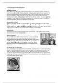
Coronary Angioplasty – force wire through femoral artery an let it travel up to
coronary arteries, insert dye and use dye to see location of narrowing. Once located,
put in a stent or balloon to reduce narrowing.
Angina Pectoris – infection of throat and discomfort in chest. Associated with myocardial
ischaemia due to reduced flow in coronary artery.
Coronary Functional Anatomy
During systole the subendocardial vessels are compressed due to increased
intraventricular pressure.
Most myocardial perfusion occurs during diastole when the subendocardial vessels
remain patent (open).
Flow never comes to 0 in right coronary artery as right ventricular pressure is always
lower than left ventricular pressure.
Most flow in coronary arteries is during diastole as they are squeezed during systole.
MYOCARDIAL ISCHAEMIA
Imbalance between oxygen demand and supply.
Increased oxygen demand = NA release.
Decreased oxygen supply = due to vasoconstriction and you get the diversion of
blood in post-prandial angina (from heart to stomach).
IHD (Ischaemic Heart Disease) – leads to stable angina – if it ruptures, get thrombus and can
lead to unstable angina.
Features of Angina
SITE – Centre of chest down left arm (commonly).
ONSET – Strss, emotion, (pain at rest is unstable angina).
CHARACTER – Tight, strangling compression.
RADIATION – Radiates to left arm, jaw and neck.
ASSOCIATED SYMPTOMS – Nausea and sweating.
TIMING – Stable angina lasts for less than 15 mins, whereas unstable angina lasts for longer
and more frequent.
EXACERBATING/RELIEVING FACTORS – Rest (relieving) and stress/exercise (exacerbating).
SEVERITY – How severe?
DIAGNOSTICS
Get atherosclerosis in coronary artery; therefore, calcification of the plaque.
Can do a CT calcium scoring to see how much calcium – if too high then do coronary
angiography.
TREATMENT
CABG (bypass stenosis by introducing artery to raise blood normal blood flow).
Lifestyle (weight loss and by reducing smoking).
Medication (Aspirin, Statins, Beta-blockers).
Coronary Angioplasty – force wire through femoral artery an let it travel up to
coronary arteries, insert dye and use dye to see location of narrowing. Once located,
put in a stent or balloon to reduce narrowing.