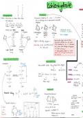
Chapter 9: Muscles
Vidz dat might help
https://www.youtube.com/watch?v=NfEJUPnqxk0
https://www.youtube.com/watch?v=f_tZne9ON7c
https://www.youtube.com/watch?v=Vs0tZV35_pw
https://www.youtube.com/watch?v=MZJ6kTKDFmw
Gamay rna kapoy tanaw sa uban hohoho
A. Skeletal Muscle
-attached to bone; its contraction is responsible for supporting and moving the
skeleton
-voluntary control
-striated muscle (have alternating light and dark bands; stripes)
-skeletal muscle cell is aka muscle fiber; each skeletal muscle fiber:
>has an elongated shape
>has multiple nuclei
>Is extremely large
>is formed during the fusion of undifferentiated, mononucleated cells (myoblasts)
into single, cylindrical, multinucleated cells
>differentiation is completed around time of birth, but continue to increase in size
to adulthood
>each participates in regulation of gene expression and protein synthesis
-when skeletal muscle fibers are damaged (ex. injury), repair involves satellite cells
*Satellite cells
-undifferentiated stem cells
-located between the plasma membrane and surrounding basement membrane
-in response to strain or injury, they become active and undergo mitotic proliferation
(daughter cells differentiate into myoblasts that can either fuse together to form new
fibers or fuse with stressed or damaged muscle fibers to repair them)
*Hypertrophy
-an increase in size of muscle
-satellite cell-mediated compensation for loss of muscle tissue
-occurs through a combination of hypertrophy of existing fibers, splitting of existing
fibers, and satellite cell proliferation, differentiation, and fusion
*Tendons
-by bundles of connective tissue consisting of collagen fibers
-attaching skeletal muscles to bones
*Filaments
-part of the bundles of myofibrils
A. Thick filaments
-composed almost entirely of the protein myosin
*Myosin
-composed of two large polypeptide heavy chains and four smaller light chains
-these polypeptides consist of : 2 globular heads and a long tail
a.2 heads
, - extend out to the sides, forming cross-bridges (which make contact
with the thin filament and exert force during muscle contraction)
-contains two binding sites: one for attaching to the thin filament and
one for ATP (the ATP binding site also functions as an enzyme called
myosin-ATPase, that hydrolyzes the bound ATP, harnessing its energy
for contraction)
b.tail
-formed by the two intertwined heavy chains
-lies along the axis of the thick filament
B. Thin filaments
-principally composed of the protein actin, troponin and tropomyosin
*Actin
-Each actin molecule contains a binding site for myosin
-globular protein makes up two intertwined, helical chains
*Troponin and tropomyosin
-functions in regulating contraction
troponin
-interacts with both actin and tropomyosin
-composed of three subunits designated by the letters I (inhibitory),
T (tropomyosin-binding) and C (Ca21-binding)
-troponin molecules bind to each molecule of tropomyosin and
regulates the access to myosin-binding sites on the actin monomers in
contact with that tropomyosin
tropomyosin
- a rod-shaped molecule composed of two intertwined polypeptides
-tropomyosin molecules are (1) arranged end to end along the actin
thin filament, are (2) partially cover the myosin-binding site on each
actin monomer (thereby preventing the cross-bridges from making
contact with actin), and is (3) held in
this blocking position by the smaller globular protein, troponin
! resting muscle fiber: troponin and tropomyosin cooperatively block
the interaction of cross-bridges with the thin filament
*Sarcomeres
-one unit of a repeating pattern of thick & thin filaments arranged in an orderly, parallel
manner
-each sarcomere contains two sets of thin filaments, one at each end (first end is
anchored to the Z line & the second end overlaps a portion of the thick filaments)
*Z line
-a network of interconnecting proteins
-two successive Z lines define the limits of one sarcomere
Bands:
A. A band
-wide, dark band
-where thick filaments are located (in the middle of each sarcomere)
Two additional bands are present in the A-band region of each sarcomere:
a.1 H zone