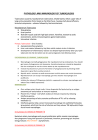
PATHOLOGY AND IMMUNOLOGY OF TUBERCULOSIS
Tuberculosis caused by mycobacterium tuberculosis, inhaled bacillus infects upper lobe of
lungs and a granuloma forms known as the Ghon focus. Causing small pleural effusions,
bronchial compression – wheeze followed by late bronchiectasis.
Mycobacterium Tuberculosis
Slow growing
Gram positive
Wall with mycolic acid with high lipid content; therefore, resistant to acidic
environments. (Limits immunoresponse against tuberculosis).
Intracellular infection
Primary Tuberculosis – (first 3 weeks)
Asymptomatic/few symptoms
Fever and malaise followed by tiny fibro calcific nodule at site of infection.
Development of inflammatory reaction via delayed hypersensitivity when you inject
tuberculin into the skin which can be used a diagnostic test (Mantoux test).
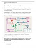
PATHOGENESIS OF PRIMARY TUBERCULOSIS
Macrophages normally phagocytose the mycobacterium by endocytosis. You should
get fusion of phagosome with lysosome; therefore bacterium should be degraded,
but this is delayed for the first three weeks by the mycobacterium.
Instead mycobacterium prevents formation of phagolysosome by blocking CA20
dependent signal that would promote it.
Mycolic acid is resistant to acidic environment and the waxy coat resists lysosomes.
Mycobacterium can escape macrophage, get into alveolar macrophages and
proliferate.
Inhibits the release of IFN-gamma (which is supposed to activate macrophages and
induce Class II MHC expression).
IMMUNOLOGY
Once antigen enters lymph node, it may phagocyte the bacterium by an antigen
presenting cell and produce an immune response.
Present it to T-helper 1 cell which evokes an immune response by releasing
interferon-gamma.
Antigen presenting cell also promotes proliferation of Th1 cell by releasing
interleukin 12.
Interferon-gamma helps convert monocyte/macrophage into epitheliod histiocytes
(granuloma), which limit the site of infection and they release TNF-alpha which helps
recruit more macrophages.
(In Summary) 0-3 WEEKS OF PRIMARY TB
Bacterium enters macrophages and you get proliferation within alveolar macrophages.
Also phagosome fusing with lysosome is prevented; therefore, preventing lytic enzymes
being produced. BACTEREMIA – asymptomatic.
(In Summary) 4-6 WEEKS OF PRIMARY TB
Tuberculosis caused by mycobacterium tuberculosis, inhaled bacillus infects upper lobe of
lungs and a granuloma forms known as the Ghon focus. Causing small pleural effusions,
bronchial compression – wheeze followed by late bronchiectasis.
Mycobacterium Tuberculosis
Slow growing
Gram positive
Wall with mycolic acid with high lipid content; therefore, resistant to acidic
environments. (Limits immunoresponse against tuberculosis).
Intracellular infection
Primary Tuberculosis – (first 3 weeks)
Asymptomatic/few symptoms
Fever and malaise followed by tiny fibro calcific nodule at site of infection.
Development of inflammatory reaction via delayed hypersensitivity when you inject
tuberculin into the skin which can be used a diagnostic test (Mantoux test).
PATHOGENESIS OF PRIMARY TUBERCULOSIS
Macrophages normally phagocytose the mycobacterium by endocytosis. You should
get fusion of phagosome with lysosome; therefore bacterium should be degraded,
but this is delayed for the first three weeks by the mycobacterium.
Instead mycobacterium prevents formation of phagolysosome by blocking CA20
dependent signal that would promote it.
Mycolic acid is resistant to acidic environment and the waxy coat resists lysosomes.
Mycobacterium can escape macrophage, get into alveolar macrophages and
proliferate.
Inhibits the release of IFN-gamma (which is supposed to activate macrophages and
induce Class II MHC expression).
IMMUNOLOGY
Once antigen enters lymph node, it may phagocyte the bacterium by an antigen
presenting cell and produce an immune response.
Present it to T-helper 1 cell which evokes an immune response by releasing
interferon-gamma.
Antigen presenting cell also promotes proliferation of Th1 cell by releasing
interleukin 12.
Interferon-gamma helps convert monocyte/macrophage into epitheliod histiocytes
(granuloma), which limit the site of infection and they release TNF-alpha which helps
recruit more macrophages.
(In Summary) 0-3 WEEKS OF PRIMARY TB
Bacterium enters macrophages and you get proliferation within alveolar macrophages.
Also phagosome fusing with lysosome is prevented; therefore, preventing lytic enzymes
being produced. BACTEREMIA – asymptomatic.
(In Summary) 4-6 WEEKS OF PRIMARY TB