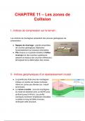
HESI Med Surg Respiratory
HESI Med Surg Respiratory How does fever cause dehydration? D/t excessive fluid loss from diaphoresis. Increased temperature also increased metabolism and the demand for O2. Clients at high risk for pneumonia: ALOC, depressed or absent gag and cough reflex, susceptible to aspirating oropharyngeal secretions, including alcoholics, anesthetized patients, brain injury, drug overdose, stroke victim, immunocompromised, immobile, cigarette smokers, other neuro disorders, debilitated by accumulated lung secretions. Nursing assessment and S/SX of pneumonia: Tachypnea- shallow respirations, often with use of accessory muscles, abrupt onset of fever with shaking and chills, productive cough with pleuritic pain, rapid bounding pulse. Elevated WBC's, pain and dullness to percussion over affected lung area, bronchial breath sounds, crackles, chest radiograph indicative of infiltrates. ABG might indicate hypoxemia. S/SX of pneumonia in older adults: Confusion, lethargy, malaise, anorexia, rapid RR, tachypnea. Drugs for pneumonia: 1. Penicillins 2. Semisynthetics 3. Penicillins and Combos: Ampicillin, Unasyn, Zosyn. 4. Tetracylcines: Tetraclycine HCL, Vibramycin, Minocin 5. Aminoglycosides: Tobramycin, Gentamicin. 6. Misc: Vancomycin, Flagyl. 7. Cephlaosporins: Kefzol, Keflex, Manol, Cefzil, Suprax. Bronchial breath sounds in pneumonia: Heard over areas of density or consolidation. Sound waves are easily transmitted over consolidated tissues. Hydration in pneumonia: Thins out the mucus trapped in the bronchioles and alveoli, facilitating expectoration. Is essential for client experiencing fever b/c diaphoresis. Is important because 300-400mL of fluid is lost in the lungs daily by evaporation. Client should also have fluids up to 3L/day Early signs of cerebral hypoxia: Irritability and restlessness. The client's brain is not receiving enough O2. Pneumonia Preventives: Older adults: get flu shot, pneumonia immunizations; avoid sources of infection and indoor pollutants- dust, smoke, aerosols; no smoking. Immunosuppressed and debilitated persons: infection avoidance, sensible nutrition, adequate intake, balance of rest and activity. Comotose and immobile patients: elevate HOB to feed and for 1 hr after feeding; frequently turning. Patients with functional or anatomic asplenia: Fku and pneumonia immunizations. Chronic Bronchitis assessment: "Blue bloaters". Generalized cyanosis, right sided heart failure, distended neck veins , crackles, expiratory wheezes. Emphysema assessment: "Pink puffers", barrell chest, pursed lip breathers, distant quiet breath sounds, wheezes, pulmonary blebs on radiograph- air trappings can explode next to each other and cause pneumothorax. Asthma assessment: Dyspnea, wheezing, chest tightness, assess precipitating factors, medication history. Compensation with COPD: ABG's are altered. As COPD worsens, the amount of O2 in blood decreased- hypoxemia, and the amount of CO2 in the blood increased-hypercapnia. This caused chronic respiratory acidosis-> increased arterial CO2 (PCO2) which results metabolic alkalosis- increased arterial bicarbonate, as compensation. Not all clients with COPD are CO2 retainers, even when hypoxemia is present because CO2 diffuses more easily across lung membranes than O2. Advanced emphysema: b/c alveoli are more affected, hypercarbia is a problem rather than bronchitis- where airways are affected. Baseline date obtained for these patients- imperative. Nursing assessment for COPD: changes in breathing pattern - increase in rate and increase in depth, use of accessory muscles-barrel chest, generalized cyanosis of lips, mouth, face, nail beds, cough-dry of productive, higher CO2 than average, low O2 as determined by pulse ox, decreased breath sounds, poor nutrition or weight loss, activity intolerance, coarse crackles in lung fields that usually clear after coughing, wheezing, dyspnea, orthopnea, anxiety. How can productive cough and comfort be fascinated in pt with COPD: Semi - fowler's or high fowler's, which lessens pressure on the diaphragm by abdominal organs. Gastric dissension becomes a priority in these clients because it elevates the diaphragm and inhibits full lung expansion. ABG values: pH: 7.35-7.45 PCO2: 35-45 PHCO3: 21-28 PO2: 80-100 Pink Puffer: Barrel chest is indicative of emphysema and is cause dby use of the accessory muscles to breathe. The person works harder to breathe but the amount of O2 taken in is adequate to oxygenate the tissues. Blue bloater: Insufficient oxygenation occur with chronic bronchitis and leads to generazlied cyanosis and often right signed heart failure (for pulmonae) Signs of inadequate arterial oxygenation: Cyanosis and slow cap refill >3 seconds. A chronic sign is clubbing of the finger nails and a late sign is clubbing of the fingers. Imp interventions for COPD: and health promotion: Tripod position, diaphragmatic breathing, pursed lip breathing, O2 at 1-2L/min, Small meals often, increase calories and protein but don't overfeed, pace activities to conserve energy, adequate fluid intake of 3L day. Fluids between meals rather than with- to prevent excess stomach dissension and decrease pressure on diaphragm, relaxation techniques, smoking cessation, prevention of secondary infections. Get immunizations- flu and pneumonia. Decrease caffeine d/t diuretic effect. Mechanically soft diets sometimes recommended- does not require as much chewing and digestion. Prevent secondary infections- avoid crowds, contact with people with infections disease and respiratory irritants- tobacco smoke. Supplements: Vit C for people who still smoke, Magnesium and Calcium b/c role in muscle contraction and relaxation. Routine monitoring of Mg and phosphorus- role r/t bone density- osteoporosis. DO PURPLE PAGES LATER with meds, nursing skills of respiratory client i.e. suctioning and ventilator settings P66. ... Prioritize nursing actions: CPR: ABC CAB Look and listen to COPD/respiratory patient If breath sounds are clear but the client is cyanotic and lethargic, adequate oxygenation is not occurring Key to respiratory assessment: Assessment of breath sounds as well as visualization of the client. Breath sounds are better described, not named: i.e. crackles, wheezes or high pitched whistling sound rather than rales, rhonchi etc. b/c might not mean same to each clinical professional. O2 delivery In adults: O2 must bubble through some type of water solution so it can be humidified if given >4L/min or delivered directly to the trachea. If given at 1-4 L/min or by mask or nasal prongs, the oropharynx and nasal pharynx provide adequate humidification. CA of larynx: often d/t cigarette smoking and alcohol. Assessment: Assess for color changes in mouth or tongue, hoarseness longer than 2 weeks, MRI, dyspnea, dysphagia, cough, hemoptysis, weight loss, pain radiating to ear, enlarged cervical nodes, halitosis. Earliest sign of CA of larynx: Hoarseness or change in vocal quality that lasts more than 2 weeks Tongue and mouth in CA of larynx: White, dark brown, gray, or black or patchy. Tracheostomy care involves: Suctioning, inner cannula change, and apply clean dressings. How does air entering lungs get humidified? Air is humidified along the nasobronchial tree. This natural humidifying pathway is gone for the patient how has had a laryngectomy. If the air is not humidified before entering the lungs, the secretions tend to thicken and become crusty. Greatest postoperative risks for laryngectomy: Bleeding or occlusion. Biggest risk time is first 24 hours. This tube has a larger lumen than tracheostomy tube but is shorter. Fear of choking in laryngectomy patients: Very real fear. They cannot cough as the could before because the glottis is gone. The the glottal stop technique to remove secretions (take a deep breath, momentarily occlude the tracheotomy tube, cough, and simultaneously remove the finger from the tube). Positive TB skin test: Induration of 10mm of greater in diameter 48-72 hours after skin test is given. BCG vaccine: Will have a positive skin test and must be evaluated with initial chest x-ray. A health hx with s/sx form may bee filled out annually until s/sx arise. When can client with TB return to work? Must have 3 negative sputum cultures TB s/sx. AIRBORNE Often asymptomatic. Symptoms include: fever with night sweats, anorexia, weight loss, malaise, fatigue, cough, hemoptysis, positive sputum culture, repeated URI's, dyspnea, pleuritic chest pain, calcification on chest x-ray . Teaching with TB: Drug therapy is usually long, >6 months, could be up to a year. It is essential that the client take the medications as prescribed for the entire time. Skipping doses or prematurely ending the drug therapy could result in a public health hazard. Teaching points with TB meds: Rifampin: can cause orange body fluids, stains contact lenses, and reduces effectiveness of birth control- should use alternative method while being tx with Rifampin. INH or Isoniazid: causes increased Dilantin levels Ethambutol: vision check before starting therapy and monthly thereafter, may have to take for 1-2 years. Teach rationale for combination drug therapy to increase complacence. Resistance develops more slowly if several anti-TB drugs are given, instead of just one. Nursing assessment of lung CA: Dry hacking cough, hoarseness, dyspnea, pain in the chest area, hemoptysis; rust colored or purulent sputum, abnormal chest radiograph, positive sputum for cytology and for pleural fluid, cough turns productive as diease progresses, diminished breath sounds, wheezing. Interventions for ung CA: Similar to those of COPD. pursed lip breathing, semi Folwer's position, relaxation techniques. Decrease pain to manageable level by administering analgesics as needed- within safety range for respiratory difficulty. Give client contrail. Keep client and family updated of impending tests and procedures. Different types of pathophysiologic conditions for nursing diagnosis ineffective breathing patterns: Inability of air sacs to fill and empty properly- emphysema and CF. Obstruction of the air passages- chronic bronchitis, asthma, and carcinoma Accumulation of fluid in the air sacs- pneumonia Respiratory muscle fstigue- COPD, pneumonia What happens if a tumor on the lung is so big you have to remove entire lobes of the lungs? When removed, large spaces are left. Chest tubes are usually not used b/c it is helpful if the mediastinal cavity, where the lung used to be, fills up with fluid. This fluid helps to prevent the shift of the remaining chest organs to fill the empty space. Upgrade to remove ads Only $3.99/month What do you do if the chest tube becomes disconnected? Do NOT clamp! Immediately place the end of the tube in a container of sterile saline of water until a new drainage system can be connected. If the chest tube is accidentally removed from the client, cover with dry sterile dressing. If an air leak is noted, tape the dressing on 3 sides only; this allows air to escape and prevents the formation of a tension pneumothroax. Notify the health care provider. NCLEX - RN content on Chest Tubes Fluctuations aka tidaling- in the fluid will occur if there is no external suction. These fluctuating movements are a good indicator that the system is intact; they should move upward with each inspiration and downward with each expiration. If fluctuations cease, check for kinked tubing, accumulation of fluid in the tubing, occlusions, or change in the client's position, because expensing lung tissue may be occluding the tube opening. When external suctioning is applied, the fluctuations cease!
Written for
- Institution
- HESI Med Surg Respiratory
- Module
- HESI Med Surg Respiratory
Document information
- Uploaded on
- January 10, 2023
- Number of pages
- 4
- Written in
- 2022/2023
- Type
- Exam (elaborations)
- Contains
- Questions & answers
Subjects
-
hesi med surg respiratory
-
hesi med surg respiratory how does fever cause dehydration dt excessive fluid loss from diaphoresis increased temperature also increased metabolism and the demand for o2
Also available in package deal