Histologie Bloed en bloedvorming 1e Ba BMW
1. Inleiding
- Vormt ongeveer 8-10% van lichaamsgewicht
- = bijzondere vorm van bindweefsel
o Amorfe matrix (=vloeibare tussenstof) = bloedplasma
o Vezels (vanuit fibrinogeen; cfr stolling)
o Cellen:
§ Erythrocyten (rode bloedcellen) < Gr erythros: rood
§ Leukocyten (witte bloedcellen) < Gr Leukos:wit
§ Thrombocyten (bloedplaatjes) < Gr thrombo: klonter
- = ‘circulerend’ weefsel
o Defensiesysteem
o Zuurstof/CO2 transport (vnl RBC)
o Vervoer nutriënten, hormonen, enzymen, vitaminen, …
o Afvoer van schadelijke stoffen naar excretieorganen
o Rol in homeostase
2. Bloedplasma
= waterige oplossing voor 10% bestaande uit opgeloste stoffen:
- anorganische zouten (0,9%)
- Organische stoffen: aminozuren, vitaminen, hormonen, glucose,…
- Plasma-eiwitten (7%):
o albuminen (belang colloid osmotische druk)
o fibrinogeen (vorming fibrine)
o Globulinen
Wanneer bloed uit circulatie à stolling
Bloedserum = plasma zonder bloedstollingsfactoren
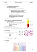
Hematocrietbepaling = verhouding # bloedcellen/totaal bloedvolume (Wright kleuring zie webtekst)
3. Bloeduitstrijkje
Routinekleuring (vb: May-Grünwald-Giemsa)
- Combinatie van kleurstoffen
o Methyleenblauw
o Azuren
o Eosine
Cellen kunnen
- basofilie (veel basische componenten is plasma)
- azurofilie, eosinofilie of neutrofilie vertonen!
4. Bloedbeeld
mens per µl bloed: 8000 -12000 leukocyten; 200000 tot 400000 bloedplaatjes; 5 -10 miljoen RBC
1
, Histologie Bloed en bloedvorming 1e Ba BMW
5. Erythrocyten (RBC)
- Meest voorkomende bloedcellen
- Levensduur is speciesafhankelijk
- Hoofdfunctie: transport zuurstof en CO2 à hemoglobine (oxy-, carbamino-) (rode kleur)
- Biconcave schijfjes voor vergroting hoge oppervlak/volume ratio
- Celmembraan verankerd met actine-bevattend cytoskelet voor behoud vd vorm
- Celmembraan bevat integrale membraaneiwitten
- Diameter (mens): 7,5 µm ;2,5 µm aan de rand; < 1 µm in het midden
- Matuur stadium heeft bij mammalia geen kern en geen andere celorganellen
- Rijpe erythrocyten ontstaan vanuit reticulocyt (zie bloedvorming).
- Soms kernfragmenten (Howell-Jolly bodies) (bij sommige diersoorten, slechts in 1% vd RBC)
5.1. Beoordelingscriteria
- RBC zijn sterk vervormbaar (cfr doorgang capillairen)
- Poikilocytosis is het voorkomen van RBC met abnormale vorm (zie dia 9)
- Eventuele hemolyse (barsten van bloedcel)
- Beschrijving op basis van:
o Diameter: microplaan – normoplaan – macroplaan – anisoplanie
o Volume: microcytair – normocytair – macrocytair
o Kleurbaarheid: hypochroom – normochroom – hyperchroom – polychroom
- Aantal RBCs
o Verhoogt à erythrocytose of polycytemie
o Verlaagt à anemie (= tekort aan Hb)
§ Te weinig of deficiënte aanmaak Hb (hypochrome anemie)
§ Tekort aan RBC (normochrome anemie)
§ Verhoogde afbraak van RBC (hemolytische anemie)
§ Microcytaire – macrocytaire – normocytaire anemie
6. Leukocyten (witte bloedcellen)
6.1. Inleiding
- 1% van totale bloedvolume
- Afweer org tg binnengedrongen vreemde materie (vb. bacteriën, parasieten, implantaten,…)
- Kunnen stromend bloed verlaten en naar het interstitium kruipen à diapedese
6.2. Indeling
Granulocyten vs agranulocyten: aanwezigheid van ‘granula’
Polymorfonucleair vs mononucleair: kernstructuur
Myeloïd vs lymfoïd: differentiatie (cfr. bloedvorming)
6.3. Granulocyten
6.3.1. Granula
1) Primaire ‘niet-specifieke’
o Lysosomen
o Azurofiel
2) Secundaire ‘specifieke’
2
1. Inleiding
- Vormt ongeveer 8-10% van lichaamsgewicht
- = bijzondere vorm van bindweefsel
o Amorfe matrix (=vloeibare tussenstof) = bloedplasma
o Vezels (vanuit fibrinogeen; cfr stolling)
o Cellen:
§ Erythrocyten (rode bloedcellen) < Gr erythros: rood
§ Leukocyten (witte bloedcellen) < Gr Leukos:wit
§ Thrombocyten (bloedplaatjes) < Gr thrombo: klonter
- = ‘circulerend’ weefsel
o Defensiesysteem
o Zuurstof/CO2 transport (vnl RBC)
o Vervoer nutriënten, hormonen, enzymen, vitaminen, …
o Afvoer van schadelijke stoffen naar excretieorganen
o Rol in homeostase
2. Bloedplasma
= waterige oplossing voor 10% bestaande uit opgeloste stoffen:
- anorganische zouten (0,9%)
- Organische stoffen: aminozuren, vitaminen, hormonen, glucose,…
- Plasma-eiwitten (7%):
o albuminen (belang colloid osmotische druk)
o fibrinogeen (vorming fibrine)
o Globulinen
Wanneer bloed uit circulatie à stolling
Bloedserum = plasma zonder bloedstollingsfactoren
Hematocrietbepaling = verhouding # bloedcellen/totaal bloedvolume (Wright kleuring zie webtekst)
3. Bloeduitstrijkje
Routinekleuring (vb: May-Grünwald-Giemsa)
- Combinatie van kleurstoffen
o Methyleenblauw
o Azuren
o Eosine
Cellen kunnen
- basofilie (veel basische componenten is plasma)
- azurofilie, eosinofilie of neutrofilie vertonen!
4. Bloedbeeld
mens per µl bloed: 8000 -12000 leukocyten; 200000 tot 400000 bloedplaatjes; 5 -10 miljoen RBC
1
, Histologie Bloed en bloedvorming 1e Ba BMW
5. Erythrocyten (RBC)
- Meest voorkomende bloedcellen
- Levensduur is speciesafhankelijk
- Hoofdfunctie: transport zuurstof en CO2 à hemoglobine (oxy-, carbamino-) (rode kleur)
- Biconcave schijfjes voor vergroting hoge oppervlak/volume ratio
- Celmembraan verankerd met actine-bevattend cytoskelet voor behoud vd vorm
- Celmembraan bevat integrale membraaneiwitten
- Diameter (mens): 7,5 µm ;2,5 µm aan de rand; < 1 µm in het midden
- Matuur stadium heeft bij mammalia geen kern en geen andere celorganellen
- Rijpe erythrocyten ontstaan vanuit reticulocyt (zie bloedvorming).
- Soms kernfragmenten (Howell-Jolly bodies) (bij sommige diersoorten, slechts in 1% vd RBC)
5.1. Beoordelingscriteria
- RBC zijn sterk vervormbaar (cfr doorgang capillairen)
- Poikilocytosis is het voorkomen van RBC met abnormale vorm (zie dia 9)
- Eventuele hemolyse (barsten van bloedcel)
- Beschrijving op basis van:
o Diameter: microplaan – normoplaan – macroplaan – anisoplanie
o Volume: microcytair – normocytair – macrocytair
o Kleurbaarheid: hypochroom – normochroom – hyperchroom – polychroom
- Aantal RBCs
o Verhoogt à erythrocytose of polycytemie
o Verlaagt à anemie (= tekort aan Hb)
§ Te weinig of deficiënte aanmaak Hb (hypochrome anemie)
§ Tekort aan RBC (normochrome anemie)
§ Verhoogde afbraak van RBC (hemolytische anemie)
§ Microcytaire – macrocytaire – normocytaire anemie
6. Leukocyten (witte bloedcellen)
6.1. Inleiding
- 1% van totale bloedvolume
- Afweer org tg binnengedrongen vreemde materie (vb. bacteriën, parasieten, implantaten,…)
- Kunnen stromend bloed verlaten en naar het interstitium kruipen à diapedese
6.2. Indeling
Granulocyten vs agranulocyten: aanwezigheid van ‘granula’
Polymorfonucleair vs mononucleair: kernstructuur
Myeloïd vs lymfoïd: differentiatie (cfr. bloedvorming)
6.3. Granulocyten
6.3.1. Granula
1) Primaire ‘niet-specifieke’
o Lysosomen
o Azurofiel
2) Secundaire ‘specifieke’
2