Oncogenes and tumour suppressor genes…Take
2
Note: it is imperative to use figures/ drawings in the essay
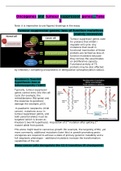
Tumour suppressor genes: loss of function mutations
(mainly recessive)
Tumour suppressor genes code
for proteins that inhibit /
regulate cell cycle. Any
mutations that result in
functional inactivation of these
proteins are termed as loss-of-
function mutations because
they remove the usual brakes
on proliferative capacity.
Functional activity of TS
proteins may be also affected
by inhibitory / competing oncoproteins or deregulation (phosphorylation status).
Knudson’s ‘two-hit’
hypothesis: loss of
heterozygosity (‘LOH’)
Typically, tumour-suppressor
genes control entry into the cell
cycle (for example, the
retinoblastoma (Rb) gene) and
the response to genotoxic
damage (for example, p53).
In paediatric neoplasms (child
cancer), mutations occur in
tumour-suppressor genes, and
both parental alleles must be
targeted (which is known as
Knudson's two-hit hypothesis). Acquisition of 2 nd mutation after getting 1st
mutant allele from parent.
This alone might lead to cancerous growth (for example, the targeting of Rb), yet
more commonly, additional mutations (later hits) in growth promoting proto-
oncogenes are required to achieve a state of primary genomic instability and a
full neoplastic phenotype – additional mutations increase the transformation
capabilities of the cell.
Many cancer cells deregulate the G1-S transition (R checkpoint):
, At this transition between G1 and S phase, cyclin E
associates with CDK2. Tumour suppressor genes
determine whether this interaction occurs and the cell
cycle proceeds. May lead to inappropriate progression
through the cell cycle.
The earliest tumour suppressors:
1. TP53 (53kDa protein)
2. Rb (retinoblastoma)
1. P53: the ‘two-faced’ security guard:-
Initially identified as a tumour specific nuclear antigen of 53kDa.
Security guard of the genome: ensures that the cell cycle proceeds only when it
says so.
2 faces: can promote cell proliferation OR cell death.
Cancers:
50% of all cancers show mutations in p53!
Certain types of cancers have more p53
mutations than others: they make up 90% of
the mutations leading to Squamous Cell
Carcinoma (SCC)
Types of p53 mutations: 80% point mutations
and 20% truncations
Antibody staining of p53 (red) shows overexpression of p53 in
Berkitt’s lymphoma compared to a normal lymph node. Overexpression of p53
protein (brown) also seen in breast cancer.
Knockout (KO) mice:
Both homozygous and heterozygous knockouts for the p53 allele have a shorter
life span than wild type individuals. Heterozygous knockouts live for a longer
than homozygous knockouts but both are predisposed to develop tumours.
Genotoxic stress stabilises p53:
Genotoxic stress includes ROS,
nutrient deprivation, hypoxia,
radio/chemotherapy and
oncoproteins.
2
Note: it is imperative to use figures/ drawings in the essay
Tumour suppressor genes: loss of function mutations
(mainly recessive)
Tumour suppressor genes code
for proteins that inhibit /
regulate cell cycle. Any
mutations that result in
functional inactivation of these
proteins are termed as loss-of-
function mutations because
they remove the usual brakes
on proliferative capacity.
Functional activity of TS
proteins may be also affected
by inhibitory / competing oncoproteins or deregulation (phosphorylation status).
Knudson’s ‘two-hit’
hypothesis: loss of
heterozygosity (‘LOH’)
Typically, tumour-suppressor
genes control entry into the cell
cycle (for example, the
retinoblastoma (Rb) gene) and
the response to genotoxic
damage (for example, p53).
In paediatric neoplasms (child
cancer), mutations occur in
tumour-suppressor genes, and
both parental alleles must be
targeted (which is known as
Knudson's two-hit hypothesis). Acquisition of 2 nd mutation after getting 1st
mutant allele from parent.
This alone might lead to cancerous growth (for example, the targeting of Rb), yet
more commonly, additional mutations (later hits) in growth promoting proto-
oncogenes are required to achieve a state of primary genomic instability and a
full neoplastic phenotype – additional mutations increase the transformation
capabilities of the cell.
Many cancer cells deregulate the G1-S transition (R checkpoint):
, At this transition between G1 and S phase, cyclin E
associates with CDK2. Tumour suppressor genes
determine whether this interaction occurs and the cell
cycle proceeds. May lead to inappropriate progression
through the cell cycle.
The earliest tumour suppressors:
1. TP53 (53kDa protein)
2. Rb (retinoblastoma)
1. P53: the ‘two-faced’ security guard:-
Initially identified as a tumour specific nuclear antigen of 53kDa.
Security guard of the genome: ensures that the cell cycle proceeds only when it
says so.
2 faces: can promote cell proliferation OR cell death.
Cancers:
50% of all cancers show mutations in p53!
Certain types of cancers have more p53
mutations than others: they make up 90% of
the mutations leading to Squamous Cell
Carcinoma (SCC)
Types of p53 mutations: 80% point mutations
and 20% truncations
Antibody staining of p53 (red) shows overexpression of p53 in
Berkitt’s lymphoma compared to a normal lymph node. Overexpression of p53
protein (brown) also seen in breast cancer.
Knockout (KO) mice:
Both homozygous and heterozygous knockouts for the p53 allele have a shorter
life span than wild type individuals. Heterozygous knockouts live for a longer
than homozygous knockouts but both are predisposed to develop tumours.
Genotoxic stress stabilises p53:
Genotoxic stress includes ROS,
nutrient deprivation, hypoxia,
radio/chemotherapy and
oncoproteins.