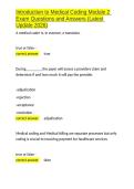
Anatomy of scapula
The scapula (Latin shoulder blade) is a thin bone placed
on the poSterolateral aspect of the thoracic cage. The
scapula has two surfaces, three borders, three angles,
and three processes
Side Determination :
1) The lateral or glenoid (Greek socket) angle is large
and bears the glenoid cavity.
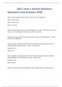
2 )The dorsal surface is convex and is divided by the
triangular Spine into the supraspinous and
infraspinous tossae. The costal Surface is occupied
by the concave subscapular fossa to fit on the convex
chest wall.
3 )The thickest lateral border runs from the glenoid
cavity above to the inferior angle below.
Features :
a)Surfaces :
1 )The costal surface or $subscapular fossa is concave and
is directed medially and forwards. It is marked by three longitudinal ridges. Another thick ridge
adjoins the lateral border. This part of the bone is almost rod-like. It acts as a lever for the action
of the serratus anterior in overhead abduction of the arm.
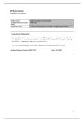
2) The dorsal surface gives attachment to the spine of scapula which divides the surface into a
smallersupraspinous fossa and a larger infraspinous fossa. The infraspinous fossae are
connected by the spinoglenoid notch,
Situated lateral to the root of the spine.
b) Borders :
1) The superior border is thin and shorter. Near the root of the coracoid process, it presents the
suprascapular notch.
2) The Lateral border is thick. At The upper end, it present the infraglenoid tubercle.
3 )The medial border is thin. It extends from the superior angle to the inferior angle.
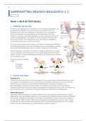
c) Angles :
1 )The superior angle is covered by the trapezius.
The scapula (Latin shoulder blade) is a thin bone placed
on the poSterolateral aspect of the thoracic cage. The
scapula has two surfaces, three borders, three angles,
and three processes
Side Determination :
1) The lateral or glenoid (Greek socket) angle is large
and bears the glenoid cavity.
2 )The dorsal surface is convex and is divided by the
triangular Spine into the supraspinous and
infraspinous tossae. The costal Surface is occupied
by the concave subscapular fossa to fit on the convex
chest wall.
3 )The thickest lateral border runs from the glenoid
cavity above to the inferior angle below.
Features :
a)Surfaces :
1 )The costal surface or $subscapular fossa is concave and
is directed medially and forwards. It is marked by three longitudinal ridges. Another thick ridge
adjoins the lateral border. This part of the bone is almost rod-like. It acts as a lever for the action
of the serratus anterior in overhead abduction of the arm.
2) The dorsal surface gives attachment to the spine of scapula which divides the surface into a
smallersupraspinous fossa and a larger infraspinous fossa. The infraspinous fossae are
connected by the spinoglenoid notch,
Situated lateral to the root of the spine.
b) Borders :
1) The superior border is thin and shorter. Near the root of the coracoid process, it presents the
suprascapular notch.
2) The Lateral border is thick. At The upper end, it present the infraglenoid tubercle.
3 )The medial border is thin. It extends from the superior angle to the inferior angle.
c) Angles :
1 )The superior angle is covered by the trapezius.