Ransley Fernandes Admin no.40202
PHYSICS ASSIGNMENT
ULTRASOUND
BACKGROUND
https://www.google.com/url?sa=i&url=https%3A%2F%2Fen.wikipedia.org%2Fwiki
%2FUltrasound&psig=AOvVaw3m_mg6H77H6o6nLUQnrGsi&ust=1576756498639000&source=images&cd=vfe
&ved=0CAIQjRxqFwoTCPjV0-mRv-YCFQAAAAAdAAAAABAE
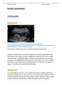
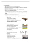
This image is of the foetus that is monitored using the ultrasound.
Humans are able to hear sound with frequency of 20 Hertz and 20,000 Hertz;
Ultrasound is any frequency above 20,000 Hertz. Ultrasound is used to image
inside the body. Ultrasound probes transducer which produces sound waves,
soundwaves are longitudinal. Ultrasound are non-invasive, so it is not harmful
therefore we use ultrasound for monitoring foetus. To advance the quality of
image, the probe can be placed inside the body.
PRODUCTION
It is not possible for humans or for machine to be able to vibrate something
more than 20,000 cycles in one second, so we use electricity and piezo- electric
crystals to produce ultrasound. Piezo electric-crystals expand or contract when
we pass electricity through them, which will induce current if vibrated.
,Ransley Fernandes Admin no.40202
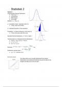
This image shows
the interior part of
transducer and the
ultrasound is
produced through
piezo-electric
crystals
https://www.google.com/url?
sa=i&source=images&cd=&ved=2ahUKEwiY1Nb3y8HmAhWOz4UKHfvKDgUQjRx6BAgBEAQ&url=https%3A%2F
%2Fwww.vaultrasound.com%2Feducational-resources%2Fultrasound-physics%2Ftransducers
%2F&psig=AOvVaw3yoZd2GcHN0ZaOib-sSQRQ&ust=1576840745284275
When we apply the current, the piezo-electric crystals vibrate and change
shape rapidly. This rapid change or vibration of the crystals produce
soundwaves that will travel outward. The alternating current will go back and
forth for more than 20,000 times in 1 second. When sound waves hit the
crystals, they release electrical currents. Hence, the same piezo-electric
crystals can be used to send and receive sound waves. The probe has a
substance that absorbs sounds to remove back reflections from the probe, and
an acoustic lens to help focus the released sound waves. The piezo-electric
crystal passes the current to the computer and creates an image.
INSIDE THE BODY
Some transducers are placed internally, to get informative and clearer image.
For example, an endorectal transducer, for the use of rectum. When
Ultrasound travels into the body, at each boundary (fat/tissue) some waves
, Ransley Fernandes Admin no.40202
will reflect, and others will keep moving; the waves that enter the body do not
have any effect on those leaving as they pass through each other. The
computer will than produce an image.
USES
Ultrasound is used to lead procedures such as needle biopsies, in which
the needles are used to demonstrate cells from an unusual area for
testing in laboratory.
Ultrasound is also used to image the breasts and guide the biopsy of
breast cancer
Doppler Ultrasound images can help the physician to observe and assess
the blockages for the flow of blood.
Ultrasound detect if there are any narrow vessels by giving an image.
Through ultrasound we can see if the person has tumours and
congenital vascular malformation.
Ultrasound is used to monitor the foetus.
It is also used to see if the person has kidney or liver stone and other
disease.
It also detects bleeding around the heart.
ADVANTAGES
Ultrasound is extremely safe, does not use radiation.
Ultrasound is really fast; the patient can know the problem or pain
he/she are going through.
It can also do by nurse, if there is an emergency.
Ultrasound gives a live image, such as, the image of blood flowing or
heart valves.
Ultrasound is widely available; it is easy to use and are lesson expensive
than other imaging methods.
Ultrasound is helpful in recognising the nerve and to allow precise
guidance of a needle for injection.
Ultrasound can capture images of soft tissues which we cannot see with
the use of X-ray
PHYSICS ASSIGNMENT
ULTRASOUND
BACKGROUND
https://www.google.com/url?sa=i&url=https%3A%2F%2Fen.wikipedia.org%2Fwiki
%2FUltrasound&psig=AOvVaw3m_mg6H77H6o6nLUQnrGsi&ust=1576756498639000&source=images&cd=vfe
&ved=0CAIQjRxqFwoTCPjV0-mRv-YCFQAAAAAdAAAAABAE
This image is of the foetus that is monitored using the ultrasound.
Humans are able to hear sound with frequency of 20 Hertz and 20,000 Hertz;
Ultrasound is any frequency above 20,000 Hertz. Ultrasound is used to image
inside the body. Ultrasound probes transducer which produces sound waves,
soundwaves are longitudinal. Ultrasound are non-invasive, so it is not harmful
therefore we use ultrasound for monitoring foetus. To advance the quality of
image, the probe can be placed inside the body.
PRODUCTION
It is not possible for humans or for machine to be able to vibrate something
more than 20,000 cycles in one second, so we use electricity and piezo- electric
crystals to produce ultrasound. Piezo electric-crystals expand or contract when
we pass electricity through them, which will induce current if vibrated.
,Ransley Fernandes Admin no.40202
This image shows
the interior part of
transducer and the
ultrasound is
produced through
piezo-electric
crystals
https://www.google.com/url?
sa=i&source=images&cd=&ved=2ahUKEwiY1Nb3y8HmAhWOz4UKHfvKDgUQjRx6BAgBEAQ&url=https%3A%2F
%2Fwww.vaultrasound.com%2Feducational-resources%2Fultrasound-physics%2Ftransducers
%2F&psig=AOvVaw3yoZd2GcHN0ZaOib-sSQRQ&ust=1576840745284275
When we apply the current, the piezo-electric crystals vibrate and change
shape rapidly. This rapid change or vibration of the crystals produce
soundwaves that will travel outward. The alternating current will go back and
forth for more than 20,000 times in 1 second. When sound waves hit the
crystals, they release electrical currents. Hence, the same piezo-electric
crystals can be used to send and receive sound waves. The probe has a
substance that absorbs sounds to remove back reflections from the probe, and
an acoustic lens to help focus the released sound waves. The piezo-electric
crystal passes the current to the computer and creates an image.
INSIDE THE BODY
Some transducers are placed internally, to get informative and clearer image.
For example, an endorectal transducer, for the use of rectum. When
Ultrasound travels into the body, at each boundary (fat/tissue) some waves
, Ransley Fernandes Admin no.40202
will reflect, and others will keep moving; the waves that enter the body do not
have any effect on those leaving as they pass through each other. The
computer will than produce an image.
USES
Ultrasound is used to lead procedures such as needle biopsies, in which
the needles are used to demonstrate cells from an unusual area for
testing in laboratory.
Ultrasound is also used to image the breasts and guide the biopsy of
breast cancer
Doppler Ultrasound images can help the physician to observe and assess
the blockages for the flow of blood.
Ultrasound detect if there are any narrow vessels by giving an image.
Through ultrasound we can see if the person has tumours and
congenital vascular malformation.
Ultrasound is used to monitor the foetus.
It is also used to see if the person has kidney or liver stone and other
disease.
It also detects bleeding around the heart.
ADVANTAGES
Ultrasound is extremely safe, does not use radiation.
Ultrasound is really fast; the patient can know the problem or pain
he/she are going through.
It can also do by nurse, if there is an emergency.
Ultrasound gives a live image, such as, the image of blood flowing or
heart valves.
Ultrasound is widely available; it is easy to use and are lesson expensive
than other imaging methods.
Ultrasound is helpful in recognising the nerve and to allow precise
guidance of a needle for injection.
Ultrasound can capture images of soft tissues which we cannot see with
the use of X-ray