Veronika Nikolaeva, F5 inroductory work
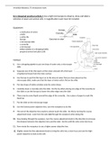
8.2.1 Required practical activity 1 Use a light microscope to observe, draw and label a
selection of plant and animal cells. A magnification scale must be included.
Equipment:
- a small piece of onion
- a knife
- a white tile
- forceps
- a microscope slide
- a coverslip
- a microscope
- iodine solution in a dropping bottle
- prepared animal and plant cells
Method:
1. Use a dropping pipette to put one drop of water onto a microscope
slide
2. Separate one of the thin layers of the onion and peel off a thin layer
of epidermal tissue from the inner surface
3. Use forceps to put this thin layer on to the drop of water that you have placed on the
microscope slide, make sure that the layer of onion cells is flat on the slide
4. Put two drops of iodine solution onto the onion tissue
5. Carefully lower a coverslip onto the slide. Do this by either placing one edge of the coverslip on
the slide or use the forceps to lower the other edge onto the slide
6. There may be some liquid around the edge of the coverslip. Use a piece of paper to soak this
liquid up
7. Put the slide on the microscope stage
8. Use the lowest power objective lens, turn the nosepiece to do this
9. The end of the objective lens needs to almost touch the slide. Do this by turning the coarse
adjustment knob. Look from the side (not through the eyepiece) when doing this
10. Now looking through the eyepiece, turn the coarse adjustment knob in the direction to increase
the distance between the objective lens and the slide. Do this until the cells come into focus
11. Now rotate the nosepiece to use a higher power objective lens
12. Slightly rotate the fine adjustment knob to bring the cells into a clear focus and use the high-
power objective to look at the cells
, Veronika Nikolaeva, F5 inroductory work
13. Make a clear, labelled drawing of some of these cells. Make sure that you draw and label any
component parts of the cell
14. Write the magnification underneath your drawing
Control variables:
- The thickness of the specimen
- Amount of iodine solution used
- Amount of water used
- The lens used
Expected results:
An enlarged picture of the structure of an onion cell, with defined nucleus, cytoplasm and vacuole
8.2.2 Required practical activity 2 (biology only)
Investigate the effect of antiseptics or antibiotics on bacterial growth using agar plates and
measuring zones of inhibition.
Equipment:
- a nutrient agar plate
- a culture of bacteria (E-coli)
- a Bunsen burner
- a glass spreader
- a heatproof mat
- filter paper discs
- three antiseptics (such as mouthwash, TCP, and antiseptic cream)
- disinfectant bench spray
- 1% VirKon disinfectant
- forceps
- clear tape
- hand wash
- a wax pencil
- access to an incubator (set to 25oC)
Method:
1. Spray the bench where you are working with disinfectant spray. Then wipe with paper towels
2. Mark the underneath of a nutrient agar plate (not the lid) with the wax pencil