HET TARSAAL
TUNNELSYNDROOM
a.Definitie
- Het is een zenuwinklemming van de N. tibialis posterior of zijn terminale takken* in de fibro-
osseuze tunnel onder het flexor retinaculum aan de mediale zijde van de enkel
(*medial/lateral plantar and calcaneal nerves = distal TTS dus meestal de laterale
plantaire zenuwtak)
- Andere mogelijke inklemmingsplaatsen:
o Mediale / laterale / intermediaire fasciale septa
b.Anatomie
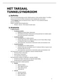
- N. tibialis posterior
o N. calcaneus medialis
Aftakking net boven flexor retinaculum
Het doorboort het flexor retinaculum om het posterieure en mediale aspect
van de hiel sensorisch te bezenuwen
o N. plantaris medialis:
Eindaftakking uit N. tibialis posterior
Passeert diep onder de M. abductor hallicus en de M. flexor hallicus longus
en verdeelt zich dan in 3 Nn. Digitalis
Levert autonome, sensorische en motorische vezels aan de plantaire voet
o N. plantaris lateralis:
Eindaftakking uit N. tibialis posterior
Passeert direct door de spierbuik van de M. abductor hallicus en gaat zo naar
de laterale zijde van de voet
Levert autonome, sensorische en motorische vezels aan de plantaire voet
- Veel anatomische variaties tov de splitsing N. plantaris medialis/lateralis
o 55% boven flexor retinaculum
o 30% thv flexor retinaculum
o 15% onder flexor retinaculum
- Veel anatomische variaties tov splitsing N. calcaneus medialis
o Vertakt boven flexor retinaculum en loopt dan oppervlakkig
o Oorsprong uit de N. plantaris lateralis (25%)
- Motorische innervatie N. tibialis posterior
o Mediaal
Abductor hallucis
Flexor digitorum brevis
Flexor hallicus brevis
Lumbricalis (I)
o Lateraal
Quadratus plantae
Flexor digiti minimi
Adductor hallicus
1
, Interossei
Lumbricalis (II-IV)
Abductor digiti minimi
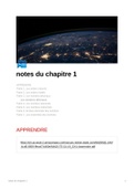
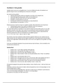
- Fibro-osseuze tunnel = tarsale tunnel
o Grenzen
Mediale malleolus (superieure rand)
Tibia (voorste rand)
Posterieur aspect van de talus (posterieure rand)
Calcaneus (laterale rand)
Abductor hallucis longis (inferieure rand)
Flexor retinaculum (mediale boord)
o Inhoud ( ‘T D A V N H’ )
M. tibialis posterior
M. flexor digitorum longus
A. tibialis
V. tibialis
N. tibialis
M. flexor hallicus longus
c. Pathofysiologie
- Compressie en/of inflammatie
- Specifieke oorzaak in 60-80% van de gevallen
- Oorzaken ingedeeld in intrinsiek, extrinsiek of combinatie van beide factoren die druk in
de tunnel kunnen verhogen
INTRINSIEK EXTRINSIEK
- Osteofyten - Trauma
- Hypertroof retinaculum - Nauw aansluitende schoenen
- Hypertofe flexor hallicus brevis - Achtervoet varus of valgus
- Lipoma - Oedeem van onderste lidmaat
- Tumor - Systemische inflammatoire
- Ganglion cysten artropathie
- Veneuze spataderen - Mucolipidose
- Pseudoaneurismen - Diabetes
- Accesory muscles - Iatrogene oorzaken
- Vasculaire leyomiomas
- Andere ruimte innemende laesies
- Distaal tarsaal tunnel syndroom = compressie van de takken van de N. tibialis
o De N. plantaris lateralis is het meest betrokken
o Combinatie van de 3 takken is ook mogelijk
o De meest voorkomende oorzaak is tractie neuritis (traumatisch)
- Risicofactor
o Bij hardlopers & voetballers: repetitieve stress en hyperpronatie geassocieerd met
slecht lopende mechanica
(puur etiologisch of enige bijdragende factor van hyperpronatie is een kwestie van
discussie)
2
TUNNELSYNDROOM
a.Definitie
- Het is een zenuwinklemming van de N. tibialis posterior of zijn terminale takken* in de fibro-
osseuze tunnel onder het flexor retinaculum aan de mediale zijde van de enkel
(*medial/lateral plantar and calcaneal nerves = distal TTS dus meestal de laterale
plantaire zenuwtak)
- Andere mogelijke inklemmingsplaatsen:
o Mediale / laterale / intermediaire fasciale septa
b.Anatomie
- N. tibialis posterior
o N. calcaneus medialis
Aftakking net boven flexor retinaculum
Het doorboort het flexor retinaculum om het posterieure en mediale aspect
van de hiel sensorisch te bezenuwen
o N. plantaris medialis:
Eindaftakking uit N. tibialis posterior
Passeert diep onder de M. abductor hallicus en de M. flexor hallicus longus
en verdeelt zich dan in 3 Nn. Digitalis
Levert autonome, sensorische en motorische vezels aan de plantaire voet
o N. plantaris lateralis:
Eindaftakking uit N. tibialis posterior
Passeert direct door de spierbuik van de M. abductor hallicus en gaat zo naar
de laterale zijde van de voet
Levert autonome, sensorische en motorische vezels aan de plantaire voet
- Veel anatomische variaties tov de splitsing N. plantaris medialis/lateralis
o 55% boven flexor retinaculum
o 30% thv flexor retinaculum
o 15% onder flexor retinaculum
- Veel anatomische variaties tov splitsing N. calcaneus medialis
o Vertakt boven flexor retinaculum en loopt dan oppervlakkig
o Oorsprong uit de N. plantaris lateralis (25%)
- Motorische innervatie N. tibialis posterior
o Mediaal
Abductor hallucis
Flexor digitorum brevis
Flexor hallicus brevis
Lumbricalis (I)
o Lateraal
Quadratus plantae
Flexor digiti minimi
Adductor hallicus
1
, Interossei
Lumbricalis (II-IV)
Abductor digiti minimi
- Fibro-osseuze tunnel = tarsale tunnel
o Grenzen
Mediale malleolus (superieure rand)
Tibia (voorste rand)
Posterieur aspect van de talus (posterieure rand)
Calcaneus (laterale rand)
Abductor hallucis longis (inferieure rand)
Flexor retinaculum (mediale boord)
o Inhoud ( ‘T D A V N H’ )
M. tibialis posterior
M. flexor digitorum longus
A. tibialis
V. tibialis
N. tibialis
M. flexor hallicus longus
c. Pathofysiologie
- Compressie en/of inflammatie
- Specifieke oorzaak in 60-80% van de gevallen
- Oorzaken ingedeeld in intrinsiek, extrinsiek of combinatie van beide factoren die druk in
de tunnel kunnen verhogen
INTRINSIEK EXTRINSIEK
- Osteofyten - Trauma
- Hypertroof retinaculum - Nauw aansluitende schoenen
- Hypertofe flexor hallicus brevis - Achtervoet varus of valgus
- Lipoma - Oedeem van onderste lidmaat
- Tumor - Systemische inflammatoire
- Ganglion cysten artropathie
- Veneuze spataderen - Mucolipidose
- Pseudoaneurismen - Diabetes
- Accesory muscles - Iatrogene oorzaken
- Vasculaire leyomiomas
- Andere ruimte innemende laesies
- Distaal tarsaal tunnel syndroom = compressie van de takken van de N. tibialis
o De N. plantaris lateralis is het meest betrokken
o Combinatie van de 3 takken is ook mogelijk
o De meest voorkomende oorzaak is tractie neuritis (traumatisch)
- Risicofactor
o Bij hardlopers & voetballers: repetitieve stress en hyperpronatie geassocieerd met
slecht lopende mechanica
(puur etiologisch of enige bijdragende factor van hyperpronatie is een kwestie van
discussie)
2