Lecture Notes from Perception to Consciousness
Lecture 1: The Retina
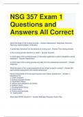
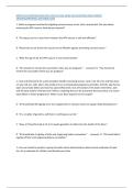
- In the retina, preprocessing happens: light passes through all the cells, but is firstly
received by the rods and cones and then send to:
o horizontal cells
o bipolar cells
o amacrine cells
o ganglion cells
- Photoreceptors: Rods (rectangular ones) and cones (pointy ones): different
wavelength sensitivity
- Rods are not involved in color vision, but are however sensitive to a specific
wavelength (greenish color)
How do rods and cones turn this into a neural signal?
- Rhodopsin (a protein): activation by light; sodium channels close.
- Different versions are sensitive to different wavelengths, which determines how
many different wavelengths you can perceive.
- Humans have 4 types: 3 for cones, 1 for rods
Retinal color blindness: absence of a cone type (Jonathan)
- Tested with “ishihara plates”
- It’s a sex-linked genetic trait; 8/100 Caucasian males
- 10x less in females!
Cone density highest in fovea (sharp vision, color vision).
- Thus, highest density of photoreceptors
- Rods mainly in parafovea (around fovea)
Light has to pass many obstacles before reaching photoreceptors
- Veins
- Vitreous body (big volume inside eye) particles “bugs”; mainly when older
Macular Degeneration: (wet or dry) blank spots and distortion in central vision; around
fovea.
- Main distinction is the loss of central vision! This is not the case in other vision-
related issues.
- Happens by loss of receptors due to accumulation of toxic products
Retinal Pigment Epithelium;
- Layer behind the photoreceptors; needs to be directly behind photoreceptors, which
is why the retinal network cells are positioned before the photoreceptors.
- Necessary for prevention of light bouncing back, so that light only activates the right
photoreceptors, not activation by reflected light.
- Cats have reflecting Pigment Epithelium (instead of absorbent), which is why they
can see better in dark light, but vision is less sharp.
, Blind Spot/Optic Disk:
- where retinal ganglion cells pass through to the optic nerve
- No photoreceptors are present
- This is because of the position of retinal ganglion cells laying on top of the
photoreceptors
Glaucoma:
- increase of pressure inside eye.
- Causes damage to nerve fibers of the retinal ganglion cells: optic nerve gets
compressed
- Treatment exists, but loss of RGC’s means lost forever.
- Hard to notice because of slow decrease of blind spots (that you don’t notice); starts
in the periphery.
Why does retina do preprocessing?
- 130.000.000 photoreceptors for each eye, but can’t all go through the 1 million
nerve fibers in optic nerve.
- So, data-compression happens. How?
Brain is mainly interested in images with high contrast between light and dark (sharp
transitions).
IMPORTANT
- Photoreceptors responds to light with Hyperpolarization; MORE NEGATIVE mV
(closing of Sodium = Na+ channels),
- Dark means de-polarization; MORE POSITIVE mV (opening of Na+ channels): graded
potential signal.
- Graded potential: means that, gradually, the lighter = more hyperpolarization (---)
The darker = more depolarization (+++)
Bipolar cells
- Positioned in between photoreceptor and retinal ganglion cells
- Necessary because photoreceptors only hyperpolarize to light, and depolarize to
dark, with only glutamate as neurotransmitter.
- (1) Sign conserving synapse = OFF bipolar cell
o Dark -> depolarization in photoreceptor -> depolarization in bipolar cell ->
action potential in ganglion cell -> IT IS DARK!
- (2) Sign inverting synapse = ON bipolar cell
o Dark -> depolarization in photoreceptor -> hyperpolarization in bipolar cell ->
NO action potential in ganglion cell -> - nothing -
o If Light: hyperpolarization in photoreceptor, depolarization in bipolar cell,
action potential in ganglion cell -> IT IS LIGHT!
o Necessary because light can otherwise not give an action potential due to
hyperpolarization when hit by light.
- They use different receptors, but same neurotransmitter
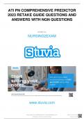
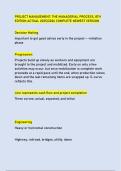
, Horizontal Cells
- Are before bipolar cells (not connected to
bipolar cells),
- Necessary for sensitivity to contrast
- Sum activity of widespread region of
photoreceptors and provide negative feedback
back on photoreceptors.
- If there is NO contrast between central and
peripheral receptive field of the group of
photoreceptors that are connected to a specific
horizontal cell, the negative feedback from the
horizontal cell works on the peripheral field, and
will depolarize the peripheral field (if
hyperpolarized by light), which evens out the
central field; resulting in no activation of the
bipolar cells.
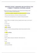
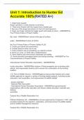
Ganglion cells only respond to contrast, not diffuse
light!
- ON-center ganglion cell
o Respond to light spot in center, dark in
surround
- OFF-center ganglion cell
o Respond to dark in center, light in
surround
- After-image (negative) is visible for both when
stimulus is taken away
- Both do not respond to diffuse light in center and surround
- Luminance by itself is not interesting to the cells, it is the contrast that we perceive
that activates action potentials; black/white = strong signal, grey/black = weaker
signal. Luminance is not necessary for us to perceive as much detail as when only
contrasts are shown
Illusions of luminance (seeing different luminance even when they are actually the same) is
because of our data compression that turns everything into contrasts
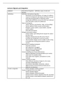
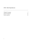
Rods
- Primarily for vision in the dark; scotopic vision
- Connect to rod-bipolar cells (different class); they receive input from multiple rods,
giving them a larger receptive field than cones!
- Different to “regular” cone-bipolar cells: they do not connect to ganglion cells
directly, but via amacrine cells
- Rods -> rod-bipolar cells -> amacrine cells -> “regular” cone-bipolar cells -> retinal
ganglion cells. ONLY for RODS
- Dark adaptation: in daylight you don’t notice their work. But when it becomes
darker, they start showing their effect. Rods are 100x more sensitive to light, thus
Lecture 1: The Retina
- In the retina, preprocessing happens: light passes through all the cells, but is firstly
received by the rods and cones and then send to:
o horizontal cells
o bipolar cells
o amacrine cells
o ganglion cells
- Photoreceptors: Rods (rectangular ones) and cones (pointy ones): different
wavelength sensitivity
- Rods are not involved in color vision, but are however sensitive to a specific
wavelength (greenish color)
How do rods and cones turn this into a neural signal?
- Rhodopsin (a protein): activation by light; sodium channels close.
- Different versions are sensitive to different wavelengths, which determines how
many different wavelengths you can perceive.
- Humans have 4 types: 3 for cones, 1 for rods
Retinal color blindness: absence of a cone type (Jonathan)
- Tested with “ishihara plates”
- It’s a sex-linked genetic trait; 8/100 Caucasian males
- 10x less in females!
Cone density highest in fovea (sharp vision, color vision).
- Thus, highest density of photoreceptors
- Rods mainly in parafovea (around fovea)
Light has to pass many obstacles before reaching photoreceptors
- Veins
- Vitreous body (big volume inside eye) particles “bugs”; mainly when older
Macular Degeneration: (wet or dry) blank spots and distortion in central vision; around
fovea.
- Main distinction is the loss of central vision! This is not the case in other vision-
related issues.
- Happens by loss of receptors due to accumulation of toxic products
Retinal Pigment Epithelium;
- Layer behind the photoreceptors; needs to be directly behind photoreceptors, which
is why the retinal network cells are positioned before the photoreceptors.
- Necessary for prevention of light bouncing back, so that light only activates the right
photoreceptors, not activation by reflected light.
- Cats have reflecting Pigment Epithelium (instead of absorbent), which is why they
can see better in dark light, but vision is less sharp.
, Blind Spot/Optic Disk:
- where retinal ganglion cells pass through to the optic nerve
- No photoreceptors are present
- This is because of the position of retinal ganglion cells laying on top of the
photoreceptors
Glaucoma:
- increase of pressure inside eye.
- Causes damage to nerve fibers of the retinal ganglion cells: optic nerve gets
compressed
- Treatment exists, but loss of RGC’s means lost forever.
- Hard to notice because of slow decrease of blind spots (that you don’t notice); starts
in the periphery.
Why does retina do preprocessing?
- 130.000.000 photoreceptors for each eye, but can’t all go through the 1 million
nerve fibers in optic nerve.
- So, data-compression happens. How?
Brain is mainly interested in images with high contrast between light and dark (sharp
transitions).
IMPORTANT
- Photoreceptors responds to light with Hyperpolarization; MORE NEGATIVE mV
(closing of Sodium = Na+ channels),
- Dark means de-polarization; MORE POSITIVE mV (opening of Na+ channels): graded
potential signal.
- Graded potential: means that, gradually, the lighter = more hyperpolarization (---)
The darker = more depolarization (+++)
Bipolar cells
- Positioned in between photoreceptor and retinal ganglion cells
- Necessary because photoreceptors only hyperpolarize to light, and depolarize to
dark, with only glutamate as neurotransmitter.
- (1) Sign conserving synapse = OFF bipolar cell
o Dark -> depolarization in photoreceptor -> depolarization in bipolar cell ->
action potential in ganglion cell -> IT IS DARK!
- (2) Sign inverting synapse = ON bipolar cell
o Dark -> depolarization in photoreceptor -> hyperpolarization in bipolar cell ->
NO action potential in ganglion cell -> - nothing -
o If Light: hyperpolarization in photoreceptor, depolarization in bipolar cell,
action potential in ganglion cell -> IT IS LIGHT!
o Necessary because light can otherwise not give an action potential due to
hyperpolarization when hit by light.
- They use different receptors, but same neurotransmitter
, Horizontal Cells
- Are before bipolar cells (not connected to
bipolar cells),
- Necessary for sensitivity to contrast
- Sum activity of widespread region of
photoreceptors and provide negative feedback
back on photoreceptors.
- If there is NO contrast between central and
peripheral receptive field of the group of
photoreceptors that are connected to a specific
horizontal cell, the negative feedback from the
horizontal cell works on the peripheral field, and
will depolarize the peripheral field (if
hyperpolarized by light), which evens out the
central field; resulting in no activation of the
bipolar cells.
Ganglion cells only respond to contrast, not diffuse
light!
- ON-center ganglion cell
o Respond to light spot in center, dark in
surround
- OFF-center ganglion cell
o Respond to dark in center, light in
surround
- After-image (negative) is visible for both when
stimulus is taken away
- Both do not respond to diffuse light in center and surround
- Luminance by itself is not interesting to the cells, it is the contrast that we perceive
that activates action potentials; black/white = strong signal, grey/black = weaker
signal. Luminance is not necessary for us to perceive as much detail as when only
contrasts are shown
Illusions of luminance (seeing different luminance even when they are actually the same) is
because of our data compression that turns everything into contrasts
Rods
- Primarily for vision in the dark; scotopic vision
- Connect to rod-bipolar cells (different class); they receive input from multiple rods,
giving them a larger receptive field than cones!
- Different to “regular” cone-bipolar cells: they do not connect to ganglion cells
directly, but via amacrine cells
- Rods -> rod-bipolar cells -> amacrine cells -> “regular” cone-bipolar cells -> retinal
ganglion cells. ONLY for RODS
- Dark adaptation: in daylight you don’t notice their work. But when it becomes
darker, they start showing their effect. Rods are 100x more sensitive to light, thus