Lecture 10: Muscles of the Forelimb
NB: Pretty much all of this is extra learning material I have added as I was interested -
you really only need to know where to locate these muscles in the body (not the origin
and insertions) and also a bit about innervation and blood supply :)
Movements
● Muscles contract to apply a force to the bone via a tendon to cause a
movement at a joint
● Revise the types of movement that can be achieved using your own bodies,
not the joint that the movement occurs about
The types of movement vary between species
● Humans have an extensive range of movement
● Cats can supinate
● Carnivores have some adduction/abduction
● Horses limited in abduction/adduction
Match the muscles to the movements at a joint
Biceps brachii Flexion at the elbow
● 2 heads of muscle
● 1 at scapula
● 1 at scapulohumeral (shoulder)
joint
● Originates at different part of the
limb
● Inserts at same tendon
● Flexion of the elbow and
shoulder joint
Quadriceps Flexion at the stifle and hip flexor
● 4 heads and 4 separate bellies
● 3 insert at the tibial tuberosity
● 1 inserts at the hip
Triceps Extension at the stifle (human knee
● 3 heads in the human equivalent)
● 4 heads in the dog (accessory
head is the 4th)
Palmar flexors Digits of the distal forelimb
Gluteals Extension of the hip
● Insert at the hip joint
Latissiumus dorsi To retract the forelimb
Supraspinatus Shoulder extension
,Pectoralis Adducts the forelimb
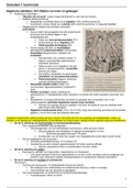
Extrinsic Musculature
● These muscles connect the limb to the trunk
● Brachiocephalicus
● Superficial pectoral
● Deep pectoral
● Osmotransversarius
● Trapexius
● Rhomboideus
● Latissimus dorsi
● Serratus ventralis
Medial
● Ventral serrate
○ Serratus ventralis
○ Holds the forelimb
● Rhomboid
○ Lies beneath the trapezius and holds the dorsal border of the scapula
close to the body
○ Holds the dorsal border of the scapula close to the trunk
○ Insertion: spine of scapula
○ Action: to elevate and abduct the forelimb
○ Innervation: accessory nerve
● Deep pectoral - superficial
○ Thorax to forelimb
○ Origin: Ventral part of sternum
○ Insertion: lesser tubercle of humerus
○ Action: when the limb is advanced and fixed in a supporting position:
to pull the trunk cranially and to extend the shoulder joint. When the
limb is not supporting weight: to draw the limb caudally and flex the
shoulder joint. To adduct the limb
○ Innervation: caudal pectoral nerves
● Superficial pectoral
, ○ Thorax to forelimb
○ Origin: the first two sternebrae
○ Insertion: the whole crest of the greater tubercle of humerus
○ Action: to adduct the limb when it is not bearing weight or to prevent
the limb from being abducted when bearing weight
○ Innervation: ventral branch of spinal nerves
Adduction of Forelimb
Lateral
● Osmotransversarius
○ In a deeper plane than the cleidocephalicus
○ Insertion: the distal end of the spine of the scapula and cranially, the
transverse process (AKA wing) of the atlas
○ Action: to advance the limb or flex the neck laterally
○ Innervation: accessory nerve
● Trapezius
○ Thin and triangular, fans out
○ Divided into cervical and thoracic parts and is separated by an
aponeurosis
○ Origin: the median raphe of the neck and the supraspinous ligament
from the level of the third cervical vertebra to the level of the ninth
thoracic vertebrae
○ Insertion: the spine of the scapula
○ Action: to elevate and abduct the forelimb
○ Innervation: accessory nerve
Aponeurosis - a sheath of pearly white tissue that takes the place of a tendon in flat
muscles having a wide area of attachment
● Latissimus dorsi
○ Large and mostly triangular
○ Lies caudal to the scapula
○ Covers most of the dorsal and some of the lateral thoracic wall
○ Origin: the thoracolumbar fascia from the spinous processes of the
lumbar and the last seven or eight thoracic vertebrae; a muscular
attachment to the last two or three ribs
NB: Pretty much all of this is extra learning material I have added as I was interested -
you really only need to know where to locate these muscles in the body (not the origin
and insertions) and also a bit about innervation and blood supply :)
Movements
● Muscles contract to apply a force to the bone via a tendon to cause a
movement at a joint
● Revise the types of movement that can be achieved using your own bodies,
not the joint that the movement occurs about
The types of movement vary between species
● Humans have an extensive range of movement
● Cats can supinate
● Carnivores have some adduction/abduction
● Horses limited in abduction/adduction
Match the muscles to the movements at a joint
Biceps brachii Flexion at the elbow
● 2 heads of muscle
● 1 at scapula
● 1 at scapulohumeral (shoulder)
joint
● Originates at different part of the
limb
● Inserts at same tendon
● Flexion of the elbow and
shoulder joint
Quadriceps Flexion at the stifle and hip flexor
● 4 heads and 4 separate bellies
● 3 insert at the tibial tuberosity
● 1 inserts at the hip
Triceps Extension at the stifle (human knee
● 3 heads in the human equivalent)
● 4 heads in the dog (accessory
head is the 4th)
Palmar flexors Digits of the distal forelimb
Gluteals Extension of the hip
● Insert at the hip joint
Latissiumus dorsi To retract the forelimb
Supraspinatus Shoulder extension
,Pectoralis Adducts the forelimb
Extrinsic Musculature
● These muscles connect the limb to the trunk
● Brachiocephalicus
● Superficial pectoral
● Deep pectoral
● Osmotransversarius
● Trapexius
● Rhomboideus
● Latissimus dorsi
● Serratus ventralis
Medial
● Ventral serrate
○ Serratus ventralis
○ Holds the forelimb
● Rhomboid
○ Lies beneath the trapezius and holds the dorsal border of the scapula
close to the body
○ Holds the dorsal border of the scapula close to the trunk
○ Insertion: spine of scapula
○ Action: to elevate and abduct the forelimb
○ Innervation: accessory nerve
● Deep pectoral - superficial
○ Thorax to forelimb
○ Origin: Ventral part of sternum
○ Insertion: lesser tubercle of humerus
○ Action: when the limb is advanced and fixed in a supporting position:
to pull the trunk cranially and to extend the shoulder joint. When the
limb is not supporting weight: to draw the limb caudally and flex the
shoulder joint. To adduct the limb
○ Innervation: caudal pectoral nerves
● Superficial pectoral
, ○ Thorax to forelimb
○ Origin: the first two sternebrae
○ Insertion: the whole crest of the greater tubercle of humerus
○ Action: to adduct the limb when it is not bearing weight or to prevent
the limb from being abducted when bearing weight
○ Innervation: ventral branch of spinal nerves
Adduction of Forelimb
Lateral
● Osmotransversarius
○ In a deeper plane than the cleidocephalicus
○ Insertion: the distal end of the spine of the scapula and cranially, the
transverse process (AKA wing) of the atlas
○ Action: to advance the limb or flex the neck laterally
○ Innervation: accessory nerve
● Trapezius
○ Thin and triangular, fans out
○ Divided into cervical and thoracic parts and is separated by an
aponeurosis
○ Origin: the median raphe of the neck and the supraspinous ligament
from the level of the third cervical vertebra to the level of the ninth
thoracic vertebrae
○ Insertion: the spine of the scapula
○ Action: to elevate and abduct the forelimb
○ Innervation: accessory nerve
Aponeurosis - a sheath of pearly white tissue that takes the place of a tendon in flat
muscles having a wide area of attachment
● Latissimus dorsi
○ Large and mostly triangular
○ Lies caudal to the scapula
○ Covers most of the dorsal and some of the lateral thoracic wall
○ Origin: the thoracolumbar fascia from the spinous processes of the
lumbar and the last seven or eight thoracic vertebrae; a muscular
attachment to the last two or three ribs