Name:
Urinary System
Structure and
Function
BTEC Applied Science
Unit 5: Principles and Applications 2 - Biology
Revision Notebook
,Functions of the urinary system
Osmoregulation
When body fluids are low
The kidney retains water in the body rather than
losing it in the urine. The result is urine with low
volume and high solute concentration.
This may be necessary on a hot day, after a lot of
physical activity or if not enough fluids are drunk.
When body fluids are high
The kidney allows more water to be lost in the urine.
The result is large volumes of dilute urine.
This is likely when the person ahs drunk a lot.
Excretion
Excretion and the origins of urea:
The urinary system is used to remove waste from the
blood. Waste includes excess salt but the main
substance removed from the body by the kidneys is
urea. Urea comes from the break down of excess
amino acids as they cannot be stored. The amino
group is formed into ammonia which is toxic. The
amino acid is combined with carbon dioxide to
produce urea. Urea will travel through the blood
stream to the kidneys where it is removed and
becomes a component of urine. The rest of the
amino acid can be converted into lipids or glucose
and used for energy.
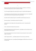
Blood pressure regulation
Two ways of regulating blood pressure
Blood pressure is increased by narrowing blood
vessels. The kidneys are one of the many organs that
can produce the hormone antigotensin 2 which
stimulates the muscles in the blood vessels to
contract making the blood vessels narrower.
The other factor that effects blood pressure is
volume of fluid. The more fluid, the higher the blood
pressure. The kidneys help regulate the amount of
fluid in the body by either retaining water if fluid
levels are low or releasing more through the urine if
fluid levels are high.
Narrow blood vessel
high pressure
Wide blood vessel low
pressure
pH homeostasis
pH balance
The blood needs to be within a narrow pH range
between 7.35 to 7.45 otherwise metabolism will be
severely affected. The urinary system helps maintain
this narrow range by excreting hydrogen ions from
the kidneys increasing the pH. Normal metabolism
releases carbon dioxide which increases the acidity of
the blood.
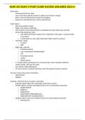
,Gross structure of the renal system:
Ureter
The ureter connects the kidney to the bladder. Its
function is to pass urine from the kidney to the
Kidney bladder.
Ureter
The bladder
The bladder is a muscular sac which is used to store
urine until it is convenient to release it from the
Bladder body. Stretch receptors in the walls detect how full it
is and stimulate the feeling of the need to pass urine.
The muscles contract to help release the urine.
Sphincter muscle
Sphincter muscle and urethra
The urine is held in the bladder by a circular muscle
called a sphincter muscle at the bottom of the
Urethra bladder. This muscle is usually tightly contracted.
When ready, the muscle can be relaxed and the urine
released to the outside via the urethra.
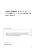
The capsule is the tough outer
Gross structure of the kidney: coating of the kidney which
Capsule protects the organ from damage.
Renal artery
The cortex is the outer layer of
the kidney between capsule and
medulla. It contains nephrons –
Cortex the active units of the kidney.
Renal vein
The medulla is the inner portion
of the kidney and is divided into
pyramids. They contain the loop
Medulla
of Henle and collecting duct parts
of the nephrons.
Ureter
The pelvis is the top part of the
ureter and is where the collecting
ducts merge together.
Pelvis
Blood supply to the kidney
The renal artery supplies blood to the kidney. The blood is needed to provide nutrients and oxygen to the kidney. However, the main reason for the
blood supply is so that the kidney can filter waste products and water from the blood.
Blood leaves the kidney via the renal vein which contains deoxygenated blood but has also had waste filtered out of it.
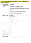
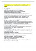
,The nephron:
The nephron function:
The function of the nephron is to filter the blood and remove unwanted waste products. Any substances needed by the body are reabsorbed from the
nephron back into the blood. The nephron also determines the amount of water that is lost in the urine or retained in the blood. This is controlled by the
hormone ADH.
Glomerulus
Efferent arteriole
Afferent arteriole
Bowman’s capsule
Distal convoluted
tubule
Proximal
convoluted tubule
Collecting duct
Descending limb
of loop of Henle
Ascending limb of
loop of Henle
Loop of Henle
Summary of processes in each section:
Structure Function summary
The Bowman’s capsule is the start of the nephron and is the location of ultrafiltration where most of the blood plasma
Bowman’s (excluding blood cells and large protein molecules) are forced out of the glomerulus and into the beginning of the
capsule nephron.
The proximal convoluted tubule is the tubule nearest to the Bowman’s capsule and is where most substances needed
Proximal by the body are reabsorbed into the blood.
convoluted tubule
The loop of Henle is a long loop which extends into the medulla of the kidney. Its function is to build up high solute
Loop of Henle (salt) concentrations in the medulla to draw water out of the nephron when needed so it can be retained by the body.
The distal convoluted tubule is the last part of the nephron before the collecting duct. It is the site of pH balance. It is
Distal convoluted also one of the two areas where water is removed from the fluid in the nephron and reabsorbed back into the blood.
tubule
The collecting duct is where a number of nephrons join together so the fluid can be conducted to the pelvis of the
Collecting duct kidney and into the ureter. It is also where water is reabsorbed from the nephron back into the blood when needed.
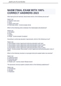
,Ultrafiltration:
Bowman’s
capsule
Afferent arteriole Efferent
arteriole
Effect of afferent arteriole being wider Constituents of filtrate:
than efferent arteriole: The filtrate contains water, salt, glucose, amino
The blood vessel to the glomerulus is wider than the acids, urea and other waste products. Blood
blood vessel leaving the glomerulus. This increases cells and proteins are too large to be squeezed
the blood pressure within the glomerulus and forces out of the blood vessels and so stay in the
most of the fluid and dissolved substances out of the blood. If kidneys are damaged protein may
blood vessels and into the Bowman’s capsule. This sometimes enter the filtrate and this is a sign of
process is called ultrafiltration. the damage.
Bowman’s capsule structure
Podocytes:
Glomerular Podocytes are foot-like cells
capillary with extensions called pedicels
which wrap around the
capillaries. The pedicels from
one podocyte interlink with the
pedicels from an adjacent
Podocyte cell podocyte. The result is small
gaps between the pedicel. These
are like the holes in a sieve and
it is through these gaps that
ultrafiltration takes place. The
Lumen gaps are too small for blood
cells or large proteins to pass, so
Pedicels these remain in the blood.
Gaps for
filtration
Capillary structure
Pedicels Gaps for
filtration
50nm Structure of glomerular capillaries:
The glomerular capillaries are similar in structure to normal
capillaries. They have a single layer of endothelial cells and an
outer layer of basement membrane. The basement membrane is
made of collagen and other fibres. It acts like filter paper only
allowing small molecules through it.
What is different about the glomerular capillaries is that they
have more pores between the individual endothelial cells than in
normal capillaries. This is to allow ultrafiltration to occur as most
substances except blood cells and large protein molecules move
through the pores during ultrafiltration.
Endothelial Basement Pores
cell membrane
,Reabsorption:
Substances reabsorbed into Substances reabsorbed into
blood in proximal convoluted blood in distal convoluted
tubule: tubule and collecting duct:
Substances such as amino acids, lipids, Most substances have already been
glucose and water are still needed by the reabsorbed from the proximal convoluted
body and so it would not be a good thing tubule. The distal convoluted tubule is for
if they were to be lost in the urine, so they the reabsorption of water.
are reabsorbed back into the blood from
the proximal convoluted tubule. There is a
very good blood supply around the
nephron to allow this. Bicarbonate ions
are also reabsorbed here leaving the
hydrogen ions to continue in the urine.
Process of reabsorption:
The sodium potassium pump: Cotransport systems: Diffusion of amino acids and
The sodium potassium pump actively Due to the low concentration of sodium in glucose:
moves 3 sodium ions out of the tubule cell the tubule cells, the sodium diffuses from Due to the tubule cells now having a
and into the surrounding tissue fluid at the filtrate in the tubule to the tubule cells higher concentration of both glucose and
the same time as pumping 2 potassium using facilitated diffusion. Cotransport is amino acids than the surrounding tissue
ions into the cell. used to move glucose and amino acids fluid, these substances both diffuse out of
This results in a low concentration of along with the sodium into the tubule the tubule cells into the surrounding
sodium ions in the tubule cell. cells. tissue fluid from where they will re-enter
the blood.
Proximal Na+ Proximal
convoluted 2K+ convoluted
tubule Tissue fluid Tissue fluid tubule
Glucose
Glucose
3Na+ Na+
Microvilli increase Amino acid
surface area for
reabsorption.
Amino acid
Build up of high salt concentrations: Action of loop of Henle (general overview):
The simple version of how the loop of Henle builds up high salt
concentrations in the medulla is as follows (this is without the counter-
current effect).
The discending limb is permeable to water and so water can leave by
osmosis. Water will leave this tube due to osmosis because the salt
concentration in the surrounding medulla is higher than the solute
concentration of the filtrate (ie. the filtrate is more dilute so water
leaves the tubule by osmosis).
Because water has left the tubule by osmosis, the salt concentration of
the filtrate at the bottom of the loop of Henle is higher than it was when
it first entered the nephron.
The cells in the walls of the ascending limb actively pump salt from the
nephron into the surrounding medulla. This pumping is against a
concentration gradient and requires energy.
The ascending limb of the loop of Henle is impermeable to water which
means that water cannot follow the salt by osmosis. If the walls were
permeable to water then water would move towards the high
concentrations of salt outside of the nephron. But this cannot happen
due to the impermeability of the ascending limb. The result is that the
filtrate at the top of the ascending limb is dilute and the medulla has
high salt concentrations.
,Counter current multiplier effect:
Purpose and effect of counter current multiplier effect:
The purpose of the counter current multiplier effect is to build up the salt concetrations in the surrounding medulla still further. The important factor is
that the ascending limb and descending limbs are next to each other with the filtrate flowing in opposite directions. This is why it is called counter-current.
This diagram represents the single effect of the loop of Henle. This is like
the first pass through of filtrate.
The result of water leaving the descending limb and salt being actively
pumped out of the ascending limb is that the salt concentration of the
filtrate is lower than the salt concentration of the filtrate that first
entered the nephron.
Because salt had been pumped out into the medulla before (as in last
picture) the medulla now has a higher salt concentration than it did. This
results in more water leaving the descending limb than would have
occurred with lower salt concentrations in the surrounding medulla.
This means that the concentration of salt that reaches the bottom of the
loop of Henle is now higher than it was before. In the last it was 400,
now it has gone up to 500 - as an example.
Because of this there is more salt available in the filtrate to be pumped
out of the ascending limb into the medulla.
The result is that the salt concentration of the surrounding medulla is
now even higher that it was before.
Because the medulla has an even higher salt concentration, even more
water leaves the descending convoluted tubule by osmosis. Water
would move by osmosis until the concentrations on the two sides are
equal if the filtrate was not moving through the nephron. Because it is
moving it does not get the chance to reach an equilibrium.
Because even more water has left the descending limb, the salt
concentrations at the bottom of the loop of Henle are now very high.
This is despite the fact that the salt concentrations of the filtrate
entering the nephron is always the same.
Due to these very large concentrations of salt in the filtrate at the
bottom of the loop of Henle, there is now even more salt available for
the ascending limb to pump out into the medulla.
,Control of water levels:
Osmoreceptors in the hypothalamus
detect that fluid levels in the blood are
low. This stimulates the pituitary gland
Low water levels to release ADH into the blood. ADH
causes distal convoluted tubule and
in blood collecting duct to become permeable
so water is reabsorbed into the blood.
Pituitary
gland
If the level of fluid in the blood is too
high, less (or no) ADH is released by
the pituitary gland. This results in
High water levels water not being able to leave the
nephron so it will be lost in the urine,
in blood rebalancing body fluid levels.
Pituitary
gland
The distal convoluted tubules and the collecting ducts are
located in the medulla of the kidney. The loop of Henle has
acted to ensure that the medulla has high salt concentrations.
High salt concentrations tend to draw water to them by
osmosis. However, if the water is unable to leave the tubule
due to it being impermeable then osmosis will not occur and
water will remain in the nephron to be eventually lost in the
urine. This is the way the kidneys work in the absence of the
hormone ADH.
If body fluid levels are low, ADH is released by the pituitary
gland and it will travel to the kidneys in the blood. ADH
changes the permeability of both the distal convoluted tubule
and the collecting duct. Now, because the walls of the tubule
are permeable to water, the high salt concentrations in the
medulla draws out the water by osmosis. The result is that
water that would have been lost in the urine is instead drawn
into the medulla where it will be reabsorbed into the blood.
This leads to lower volumes of urine which is more
concentrated and therefore darker in colour.
The salt concentration of the medulla is still high because the
actions of the loop of Henle in building salt concentrations
are not affected by ADH, they will continue regardless of the
fluid levels of the blood.
Normally, this high level of salt in the medulla would draw
water to it by osmosis. However, the water cannot leave the
distal convoluted tubules or the collecting duct because these
are usually impermeable to water. They would only become
permeable to water if there is ADH in the blood. However,
because the individual has recently had a good drink of water
the osmoreceptors in the hypothalamus to not trigger the
pituitary gland to release ADH so the two tubules remain
impermable to water.
The consequence is that water continues through the
nephron, into the collecting duct and will leave the body in
the urine. Due to the large volume of water, the urine will
have a greater volume and will be more dilute.
,How water is reabsorbed:
ADH binds to receptors
on collecting duct
cells.
Aquaporin ADH
When ADH is present in the blood it binds to receptors on the
collecting duct cells. This triggers vesicles containing
aquaporins to move towards the membrane closest to the
collecting duct.
Aquaporins are special water permeable channels which
Vesicles containing allow water to move through the membrane.
aquaporin move to the Because of the loop of Henle, the tissue fluid around the
membrane near the collecting duct has a salt concentration. This creates a low
collecting duct water potential in the tissue fluid. Now that there are pores
in the membrane, water moves by osmosis from the area of
high water potential (in the collecting duct) to an area of low
water potential (the tissue fluid of the medulla).
Consequently, excess water leaves the collecting duct and will
eventually move into the blood. The result is that the urine is
Water enters of low volume and high solute concentration.
collecting duct
cells and tissue
fluid due to high
salt concentration
of tissue fluid in
medulla.
No ADH in blood
ADH has a half-life of only about 20 minutes . It is soon
broken down. Consequently, unless the receptors in the
hypothalamus continue to signal that the body is dehydrated,
the collecting duct cells will no longer be stimulated by ADH.
The aquaporins in the membrane form back into vesicles and
move into the cell. This makes the cell membrane of the
collecting duct cells less permeable to water.
Even though the water potential of the tissue fluid is still low
due to high sodium concentrations, water cannot move into
the tissue fluid by osmosis.
As a result, the water remains in the filtrate and will be lost
out of the body as part of the urine.
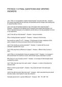
, Renin-angiotensin-aldosterone system (RAAS):
Function of RAAS:
The function of the renin-angiotensin-aldosterone system is to increase blood volume and blood pressure.
Angiotensinogen
Angiotensinogen is made and stored in the liver. When it Angiotensinogen
comes into contact with renin it is converted into
angiotensin 1.
Angiotensinogen
Renin
Renin
Renin is an enzyme that converts angiotensinogen into
angiotensin 1. It is released from cells in the afferent
arterioles, known as juxtaglomerular cells. They are Renin
triggered to release renin when blood pressure drops.
Angiotensin I
ACE ACE
Angiotensin converting enzyme (ACE)
ACE is made in the lungs and the kidneys. It is released
when blood pressure is low. When it comes into contact
with angiotensin 1, it converts it to angiotensin 2.
ACE
Angiotensin 2
Angiotensin 2 is hormone made up of only 8 amino acids.
It has a number of effects on the body which lead to an
increase in both blood pressure and blood volume. Angiotensin II
Stimulates adrenal Reabsorption of sodium
Stimulates pituitary to
cortex to produce Vasoconstriction ions in proximal
secrete ADH
aldosterone convoluted tubule
Na+
Na+ Na+
Aldosterone Vasoconstriction Sodium reabsorption ADH
Aldosterone works in the distal Angiotensin 2 causes the Reabsorption of more sodium ADH stimulates the collecting
convoluted tubules. It actively muscles of the arterioles to ions than usual causes less ducts to reabsorb more water
causes more sodium ions to be contract causing water to be lost in the urine from the nephron filtrate so less
reabsorbed from the nephron vasoconstriction. This increases because the high sodium ion is lost in the urine. This
filtrate back into the blood. This blood pressure because it concentrations in the blood increases blood volume and
increases osmosis so more reduces the space available for draw water by osmosis. This blood pressure.
water is reabsorbed increasing the blood in the cardiovascular increases blood volume and
blood volume and pressure. system. blood pressure.
Urinary System
Structure and
Function
BTEC Applied Science
Unit 5: Principles and Applications 2 - Biology
Revision Notebook
,Functions of the urinary system
Osmoregulation
When body fluids are low
The kidney retains water in the body rather than
losing it in the urine. The result is urine with low
volume and high solute concentration.
This may be necessary on a hot day, after a lot of
physical activity or if not enough fluids are drunk.
When body fluids are high
The kidney allows more water to be lost in the urine.
The result is large volumes of dilute urine.
This is likely when the person ahs drunk a lot.
Excretion
Excretion and the origins of urea:
The urinary system is used to remove waste from the
blood. Waste includes excess salt but the main
substance removed from the body by the kidneys is
urea. Urea comes from the break down of excess
amino acids as they cannot be stored. The amino
group is formed into ammonia which is toxic. The
amino acid is combined with carbon dioxide to
produce urea. Urea will travel through the blood
stream to the kidneys where it is removed and
becomes a component of urine. The rest of the
amino acid can be converted into lipids or glucose
and used for energy.
Blood pressure regulation
Two ways of regulating blood pressure
Blood pressure is increased by narrowing blood
vessels. The kidneys are one of the many organs that
can produce the hormone antigotensin 2 which
stimulates the muscles in the blood vessels to
contract making the blood vessels narrower.
The other factor that effects blood pressure is
volume of fluid. The more fluid, the higher the blood
pressure. The kidneys help regulate the amount of
fluid in the body by either retaining water if fluid
levels are low or releasing more through the urine if
fluid levels are high.
Narrow blood vessel
high pressure
Wide blood vessel low
pressure
pH homeostasis
pH balance
The blood needs to be within a narrow pH range
between 7.35 to 7.45 otherwise metabolism will be
severely affected. The urinary system helps maintain
this narrow range by excreting hydrogen ions from
the kidneys increasing the pH. Normal metabolism
releases carbon dioxide which increases the acidity of
the blood.
,Gross structure of the renal system:
Ureter
The ureter connects the kidney to the bladder. Its
function is to pass urine from the kidney to the
Kidney bladder.
Ureter
The bladder
The bladder is a muscular sac which is used to store
urine until it is convenient to release it from the
Bladder body. Stretch receptors in the walls detect how full it
is and stimulate the feeling of the need to pass urine.
The muscles contract to help release the urine.
Sphincter muscle
Sphincter muscle and urethra
The urine is held in the bladder by a circular muscle
called a sphincter muscle at the bottom of the
Urethra bladder. This muscle is usually tightly contracted.
When ready, the muscle can be relaxed and the urine
released to the outside via the urethra.
The capsule is the tough outer
Gross structure of the kidney: coating of the kidney which
Capsule protects the organ from damage.
Renal artery
The cortex is the outer layer of
the kidney between capsule and
medulla. It contains nephrons –
Cortex the active units of the kidney.
Renal vein
The medulla is the inner portion
of the kidney and is divided into
pyramids. They contain the loop
Medulla
of Henle and collecting duct parts
of the nephrons.
Ureter
The pelvis is the top part of the
ureter and is where the collecting
ducts merge together.
Pelvis
Blood supply to the kidney
The renal artery supplies blood to the kidney. The blood is needed to provide nutrients and oxygen to the kidney. However, the main reason for the
blood supply is so that the kidney can filter waste products and water from the blood.
Blood leaves the kidney via the renal vein which contains deoxygenated blood but has also had waste filtered out of it.
,The nephron:
The nephron function:
The function of the nephron is to filter the blood and remove unwanted waste products. Any substances needed by the body are reabsorbed from the
nephron back into the blood. The nephron also determines the amount of water that is lost in the urine or retained in the blood. This is controlled by the
hormone ADH.
Glomerulus
Efferent arteriole
Afferent arteriole
Bowman’s capsule
Distal convoluted
tubule
Proximal
convoluted tubule
Collecting duct
Descending limb
of loop of Henle
Ascending limb of
loop of Henle
Loop of Henle
Summary of processes in each section:
Structure Function summary
The Bowman’s capsule is the start of the nephron and is the location of ultrafiltration where most of the blood plasma
Bowman’s (excluding blood cells and large protein molecules) are forced out of the glomerulus and into the beginning of the
capsule nephron.
The proximal convoluted tubule is the tubule nearest to the Bowman’s capsule and is where most substances needed
Proximal by the body are reabsorbed into the blood.
convoluted tubule
The loop of Henle is a long loop which extends into the medulla of the kidney. Its function is to build up high solute
Loop of Henle (salt) concentrations in the medulla to draw water out of the nephron when needed so it can be retained by the body.
The distal convoluted tubule is the last part of the nephron before the collecting duct. It is the site of pH balance. It is
Distal convoluted also one of the two areas where water is removed from the fluid in the nephron and reabsorbed back into the blood.
tubule
The collecting duct is where a number of nephrons join together so the fluid can be conducted to the pelvis of the
Collecting duct kidney and into the ureter. It is also where water is reabsorbed from the nephron back into the blood when needed.
,Ultrafiltration:
Bowman’s
capsule
Afferent arteriole Efferent
arteriole
Effect of afferent arteriole being wider Constituents of filtrate:
than efferent arteriole: The filtrate contains water, salt, glucose, amino
The blood vessel to the glomerulus is wider than the acids, urea and other waste products. Blood
blood vessel leaving the glomerulus. This increases cells and proteins are too large to be squeezed
the blood pressure within the glomerulus and forces out of the blood vessels and so stay in the
most of the fluid and dissolved substances out of the blood. If kidneys are damaged protein may
blood vessels and into the Bowman’s capsule. This sometimes enter the filtrate and this is a sign of
process is called ultrafiltration. the damage.
Bowman’s capsule structure
Podocytes:
Glomerular Podocytes are foot-like cells
capillary with extensions called pedicels
which wrap around the
capillaries. The pedicels from
one podocyte interlink with the
pedicels from an adjacent
Podocyte cell podocyte. The result is small
gaps between the pedicel. These
are like the holes in a sieve and
it is through these gaps that
ultrafiltration takes place. The
Lumen gaps are too small for blood
cells or large proteins to pass, so
Pedicels these remain in the blood.
Gaps for
filtration
Capillary structure
Pedicels Gaps for
filtration
50nm Structure of glomerular capillaries:
The glomerular capillaries are similar in structure to normal
capillaries. They have a single layer of endothelial cells and an
outer layer of basement membrane. The basement membrane is
made of collagen and other fibres. It acts like filter paper only
allowing small molecules through it.
What is different about the glomerular capillaries is that they
have more pores between the individual endothelial cells than in
normal capillaries. This is to allow ultrafiltration to occur as most
substances except blood cells and large protein molecules move
through the pores during ultrafiltration.
Endothelial Basement Pores
cell membrane
,Reabsorption:
Substances reabsorbed into Substances reabsorbed into
blood in proximal convoluted blood in distal convoluted
tubule: tubule and collecting duct:
Substances such as amino acids, lipids, Most substances have already been
glucose and water are still needed by the reabsorbed from the proximal convoluted
body and so it would not be a good thing tubule. The distal convoluted tubule is for
if they were to be lost in the urine, so they the reabsorption of water.
are reabsorbed back into the blood from
the proximal convoluted tubule. There is a
very good blood supply around the
nephron to allow this. Bicarbonate ions
are also reabsorbed here leaving the
hydrogen ions to continue in the urine.
Process of reabsorption:
The sodium potassium pump: Cotransport systems: Diffusion of amino acids and
The sodium potassium pump actively Due to the low concentration of sodium in glucose:
moves 3 sodium ions out of the tubule cell the tubule cells, the sodium diffuses from Due to the tubule cells now having a
and into the surrounding tissue fluid at the filtrate in the tubule to the tubule cells higher concentration of both glucose and
the same time as pumping 2 potassium using facilitated diffusion. Cotransport is amino acids than the surrounding tissue
ions into the cell. used to move glucose and amino acids fluid, these substances both diffuse out of
This results in a low concentration of along with the sodium into the tubule the tubule cells into the surrounding
sodium ions in the tubule cell. cells. tissue fluid from where they will re-enter
the blood.
Proximal Na+ Proximal
convoluted 2K+ convoluted
tubule Tissue fluid Tissue fluid tubule
Glucose
Glucose
3Na+ Na+
Microvilli increase Amino acid
surface area for
reabsorption.
Amino acid
Build up of high salt concentrations: Action of loop of Henle (general overview):
The simple version of how the loop of Henle builds up high salt
concentrations in the medulla is as follows (this is without the counter-
current effect).
The discending limb is permeable to water and so water can leave by
osmosis. Water will leave this tube due to osmosis because the salt
concentration in the surrounding medulla is higher than the solute
concentration of the filtrate (ie. the filtrate is more dilute so water
leaves the tubule by osmosis).
Because water has left the tubule by osmosis, the salt concentration of
the filtrate at the bottom of the loop of Henle is higher than it was when
it first entered the nephron.
The cells in the walls of the ascending limb actively pump salt from the
nephron into the surrounding medulla. This pumping is against a
concentration gradient and requires energy.
The ascending limb of the loop of Henle is impermeable to water which
means that water cannot follow the salt by osmosis. If the walls were
permeable to water then water would move towards the high
concentrations of salt outside of the nephron. But this cannot happen
due to the impermeability of the ascending limb. The result is that the
filtrate at the top of the ascending limb is dilute and the medulla has
high salt concentrations.
,Counter current multiplier effect:
Purpose and effect of counter current multiplier effect:
The purpose of the counter current multiplier effect is to build up the salt concetrations in the surrounding medulla still further. The important factor is
that the ascending limb and descending limbs are next to each other with the filtrate flowing in opposite directions. This is why it is called counter-current.
This diagram represents the single effect of the loop of Henle. This is like
the first pass through of filtrate.
The result of water leaving the descending limb and salt being actively
pumped out of the ascending limb is that the salt concentration of the
filtrate is lower than the salt concentration of the filtrate that first
entered the nephron.
Because salt had been pumped out into the medulla before (as in last
picture) the medulla now has a higher salt concentration than it did. This
results in more water leaving the descending limb than would have
occurred with lower salt concentrations in the surrounding medulla.
This means that the concentration of salt that reaches the bottom of the
loop of Henle is now higher than it was before. In the last it was 400,
now it has gone up to 500 - as an example.
Because of this there is more salt available in the filtrate to be pumped
out of the ascending limb into the medulla.
The result is that the salt concentration of the surrounding medulla is
now even higher that it was before.
Because the medulla has an even higher salt concentration, even more
water leaves the descending convoluted tubule by osmosis. Water
would move by osmosis until the concentrations on the two sides are
equal if the filtrate was not moving through the nephron. Because it is
moving it does not get the chance to reach an equilibrium.
Because even more water has left the descending limb, the salt
concentrations at the bottom of the loop of Henle are now very high.
This is despite the fact that the salt concentrations of the filtrate
entering the nephron is always the same.
Due to these very large concentrations of salt in the filtrate at the
bottom of the loop of Henle, there is now even more salt available for
the ascending limb to pump out into the medulla.
,Control of water levels:
Osmoreceptors in the hypothalamus
detect that fluid levels in the blood are
low. This stimulates the pituitary gland
Low water levels to release ADH into the blood. ADH
causes distal convoluted tubule and
in blood collecting duct to become permeable
so water is reabsorbed into the blood.
Pituitary
gland
If the level of fluid in the blood is too
high, less (or no) ADH is released by
the pituitary gland. This results in
High water levels water not being able to leave the
nephron so it will be lost in the urine,
in blood rebalancing body fluid levels.
Pituitary
gland
The distal convoluted tubules and the collecting ducts are
located in the medulla of the kidney. The loop of Henle has
acted to ensure that the medulla has high salt concentrations.
High salt concentrations tend to draw water to them by
osmosis. However, if the water is unable to leave the tubule
due to it being impermeable then osmosis will not occur and
water will remain in the nephron to be eventually lost in the
urine. This is the way the kidneys work in the absence of the
hormone ADH.
If body fluid levels are low, ADH is released by the pituitary
gland and it will travel to the kidneys in the blood. ADH
changes the permeability of both the distal convoluted tubule
and the collecting duct. Now, because the walls of the tubule
are permeable to water, the high salt concentrations in the
medulla draws out the water by osmosis. The result is that
water that would have been lost in the urine is instead drawn
into the medulla where it will be reabsorbed into the blood.
This leads to lower volumes of urine which is more
concentrated and therefore darker in colour.
The salt concentration of the medulla is still high because the
actions of the loop of Henle in building salt concentrations
are not affected by ADH, they will continue regardless of the
fluid levels of the blood.
Normally, this high level of salt in the medulla would draw
water to it by osmosis. However, the water cannot leave the
distal convoluted tubules or the collecting duct because these
are usually impermeable to water. They would only become
permeable to water if there is ADH in the blood. However,
because the individual has recently had a good drink of water
the osmoreceptors in the hypothalamus to not trigger the
pituitary gland to release ADH so the two tubules remain
impermable to water.
The consequence is that water continues through the
nephron, into the collecting duct and will leave the body in
the urine. Due to the large volume of water, the urine will
have a greater volume and will be more dilute.
,How water is reabsorbed:
ADH binds to receptors
on collecting duct
cells.
Aquaporin ADH
When ADH is present in the blood it binds to receptors on the
collecting duct cells. This triggers vesicles containing
aquaporins to move towards the membrane closest to the
collecting duct.
Aquaporins are special water permeable channels which
Vesicles containing allow water to move through the membrane.
aquaporin move to the Because of the loop of Henle, the tissue fluid around the
membrane near the collecting duct has a salt concentration. This creates a low
collecting duct water potential in the tissue fluid. Now that there are pores
in the membrane, water moves by osmosis from the area of
high water potential (in the collecting duct) to an area of low
water potential (the tissue fluid of the medulla).
Consequently, excess water leaves the collecting duct and will
eventually move into the blood. The result is that the urine is
Water enters of low volume and high solute concentration.
collecting duct
cells and tissue
fluid due to high
salt concentration
of tissue fluid in
medulla.
No ADH in blood
ADH has a half-life of only about 20 minutes . It is soon
broken down. Consequently, unless the receptors in the
hypothalamus continue to signal that the body is dehydrated,
the collecting duct cells will no longer be stimulated by ADH.
The aquaporins in the membrane form back into vesicles and
move into the cell. This makes the cell membrane of the
collecting duct cells less permeable to water.
Even though the water potential of the tissue fluid is still low
due to high sodium concentrations, water cannot move into
the tissue fluid by osmosis.
As a result, the water remains in the filtrate and will be lost
out of the body as part of the urine.
, Renin-angiotensin-aldosterone system (RAAS):
Function of RAAS:
The function of the renin-angiotensin-aldosterone system is to increase blood volume and blood pressure.
Angiotensinogen
Angiotensinogen is made and stored in the liver. When it Angiotensinogen
comes into contact with renin it is converted into
angiotensin 1.
Angiotensinogen
Renin
Renin
Renin is an enzyme that converts angiotensinogen into
angiotensin 1. It is released from cells in the afferent
arterioles, known as juxtaglomerular cells. They are Renin
triggered to release renin when blood pressure drops.
Angiotensin I
ACE ACE
Angiotensin converting enzyme (ACE)
ACE is made in the lungs and the kidneys. It is released
when blood pressure is low. When it comes into contact
with angiotensin 1, it converts it to angiotensin 2.
ACE
Angiotensin 2
Angiotensin 2 is hormone made up of only 8 amino acids.
It has a number of effects on the body which lead to an
increase in both blood pressure and blood volume. Angiotensin II
Stimulates adrenal Reabsorption of sodium
Stimulates pituitary to
cortex to produce Vasoconstriction ions in proximal
secrete ADH
aldosterone convoluted tubule
Na+
Na+ Na+
Aldosterone Vasoconstriction Sodium reabsorption ADH
Aldosterone works in the distal Angiotensin 2 causes the Reabsorption of more sodium ADH stimulates the collecting
convoluted tubules. It actively muscles of the arterioles to ions than usual causes less ducts to reabsorb more water
causes more sodium ions to be contract causing water to be lost in the urine from the nephron filtrate so less
reabsorbed from the nephron vasoconstriction. This increases because the high sodium ion is lost in the urine. This
filtrate back into the blood. This blood pressure because it concentrations in the blood increases blood volume and
increases osmosis so more reduces the space available for draw water by osmosis. This blood pressure.
water is reabsorbed increasing the blood in the cardiovascular increases blood volume and
blood volume and pressure. system. blood pressure.