Anatomy lecture 7 part 2: the cerebellum
The cerebellum, which stands for “little brain”, is a structure of the central
nervous system. It has an important role in motor control, with cerebellar
dysfunction often presenting with motor signs. In particular, it is active in the
coordination of movements, as well as in motor learning.
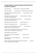
During embryonic development, the anterior portion of the neural tube forms
three parts that give rise to the brain and associated structures:
Forebrain (prosencephalon)
Midbrain (mesencephalon)
Hindbrain (rhombencephalon)
The hindbrain then divides into the metencephalon (superior) and
the myelencephalon (inferior). The cerebellum develops from the
metencephalon division.
Anatomical Location
The cerebellum is located at the back of the brain, immediately inferior to
the occipital and temporal lobes, and within the posterior cranial fossa. It
is separated from these lobes by the tentorium cerebelli, a tough layer of
dura mater.
It lies at the same level of and posterior to the pons, from which it is
separated by the fourth ventricle.
Anatomical Structure and Divisions
The cerebellum consists of two hemispheres which are connected by
the vermis, a narrow midline area. The cerebellum consists of grey matter
and white matter:
Grey matter – located on the surface of the cerebellum. It is tightly
folded, forming the cerebellar cortex.
White matter – located underneath the cerebellar cortex. Embedded
in the white matter are 4 cerebellar nuclei (the dentate, emboliform,
globose, and fastigi nuclei).
There are three ways that the cerebellum can be subdivided – anatomical
lobes, zones and functional divisions
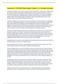
Anatomical Lobes
, There are three anatomical lobes that
can be distinguished in the
cerebellum; the anterior lobe, the
posterior lobe and the
flocculonodular lobe.
These lobes are divided by two
sulcuies – the primary
sulcus and posterolateral sulcus.
The anterior lobe extends from the level of the cerebellar peduncles
anteriorly and includes the anterior part of the superior vermis, this lobe
terminates at the primary sulcus.
From this point posteriorly and continuing along the inferior surface to the
posterolateral sulcus is the larger posterior lobe. The smallest of the lobes is
the flocculonodular lobe. It is a flattened lobe that lies between the
posterolateral sulcus (inferiorly) and the inferior medullary velum and the
cerebellar peduncles (superiorly).
Zones
There are 3 cerebellar zones. In the midline of the cerebellum is the vermis.
Either side of the vermis is the intermediate zone. Lateral to the
intermediate zone are the lateral hemispheres. There is no difference in
structure between the lateral hemispheres and intermediate zones
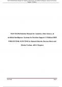
Functional Divisions
The cerebellum can also be divided
by function. There are three
functional areas of the cerebellum
– the cerebrocerebellum, the
spinocerebellum and the
vestibulocerebellum.
1. Cerebrocerebellum – the largest division, formed by the lateral
hemispheres. It is involved in planning movements and motor learning. It
The cerebellum, which stands for “little brain”, is a structure of the central
nervous system. It has an important role in motor control, with cerebellar
dysfunction often presenting with motor signs. In particular, it is active in the
coordination of movements, as well as in motor learning.
During embryonic development, the anterior portion of the neural tube forms
three parts that give rise to the brain and associated structures:
Forebrain (prosencephalon)
Midbrain (mesencephalon)
Hindbrain (rhombencephalon)
The hindbrain then divides into the metencephalon (superior) and
the myelencephalon (inferior). The cerebellum develops from the
metencephalon division.
Anatomical Location
The cerebellum is located at the back of the brain, immediately inferior to
the occipital and temporal lobes, and within the posterior cranial fossa. It
is separated from these lobes by the tentorium cerebelli, a tough layer of
dura mater.
It lies at the same level of and posterior to the pons, from which it is
separated by the fourth ventricle.
Anatomical Structure and Divisions
The cerebellum consists of two hemispheres which are connected by
the vermis, a narrow midline area. The cerebellum consists of grey matter
and white matter:
Grey matter – located on the surface of the cerebellum. It is tightly
folded, forming the cerebellar cortex.
White matter – located underneath the cerebellar cortex. Embedded
in the white matter are 4 cerebellar nuclei (the dentate, emboliform,
globose, and fastigi nuclei).
There are three ways that the cerebellum can be subdivided – anatomical
lobes, zones and functional divisions
Anatomical Lobes
, There are three anatomical lobes that
can be distinguished in the
cerebellum; the anterior lobe, the
posterior lobe and the
flocculonodular lobe.
These lobes are divided by two
sulcuies – the primary
sulcus and posterolateral sulcus.
The anterior lobe extends from the level of the cerebellar peduncles
anteriorly and includes the anterior part of the superior vermis, this lobe
terminates at the primary sulcus.
From this point posteriorly and continuing along the inferior surface to the
posterolateral sulcus is the larger posterior lobe. The smallest of the lobes is
the flocculonodular lobe. It is a flattened lobe that lies between the
posterolateral sulcus (inferiorly) and the inferior medullary velum and the
cerebellar peduncles (superiorly).
Zones
There are 3 cerebellar zones. In the midline of the cerebellum is the vermis.
Either side of the vermis is the intermediate zone. Lateral to the
intermediate zone are the lateral hemispheres. There is no difference in
structure between the lateral hemispheres and intermediate zones
Functional Divisions
The cerebellum can also be divided
by function. There are three
functional areas of the cerebellum
– the cerebrocerebellum, the
spinocerebellum and the
vestibulocerebellum.
1. Cerebrocerebellum – the largest division, formed by the lateral
hemispheres. It is involved in planning movements and motor learning. It