An Introduction to Anatomy
1. Introduction to Anatomy
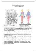
• The Anatomical Position is the
position we assume the body is in
when we discuss any aspect of
anatomy. This is the recognised
standard position to avoid confusion.
• Directional terms such as ‘the front of
the hand’ are always described with
regards to this anatomical position.
• It’s defined as looking forward,
standing upright, arms either side of
the body, palms forward and feet flat
on the floor pointing forwards. When
grounded, the bottom of the feet are
described as ventral, and the top are
described as dorsal. This is the same
with the hands.
Anterior view = view from the front.
Posterior view = view from the back.
The body can be split into sections using anatomical planes. These are imaginary lines which
can be at any angle. It enables us to look at internal cross-sectional anatomy and it’s
commonly used in clinical imaging.
Transverse Plane – splits the body into superior (upper) and inferior (lower) sections. It can
also be known as the horizontal or axial plane.
Coronal Plane – splits the body into anterior (front) and posterior (back) sections. It’s also
known as the frontal plane.
Sagittal Plane – splits the body into left and right sections. The median or mid-sagittal plane
sits directly in the midline of the body.
Surface anatomy is essential for bringing anatomy into context. It allows us to visualise the
locations of anatomical structures in relation to landmarks on the surface.
Anatomists use vertebral levels as a way of defining the location of a structure. They are
used as the vertebrate are only the only anatomical structures that are consistent between
the thorax, abdomen, and pelvis.
,The vertebrate are divided into four sections and are numbered from top to bottom.
• There are seven Cervical
vertebrate termed C1-C7.
• There are twelve Thoracic
vertebrate termed T1-T12.
• There are five Lumbar
vertebrate termed L1-L5.
• There are five Sacral
vertebrate termed S1-S5.
• There are also Coccygeal
vertebrate.
The human body can be split up into body regions. Within these regions there will be
various structures from different body systems. Having these regions allows anatomists to
compartmentalise the body.
Head and Neck – The head contains the brain and sensory organs and houses parts of the
respiratory and gastrointestinal tracts. The neck acts a passageway between the head and
thorax and contains blood vessels coming from the head and spinal cord towards more
inferior structures. Two important structures in the neck are the larynx and the oesophagus.
Trunk – This is the central mass of the human body that can be split into three regions:
thoracic, abdominal, and pelvic cavities.
Thorax – This contains the heart, lungs, and great vessels. The boundaries of the thorax are
largely made up of the thoracic cage and thoracic wall muscles. The thoracic cavity is the
space within the thoracic cage and can be split into pleural cavities and the mediastinum.
Abdomen – This extends from the diaphragm superiorly to the inguinal ligaments and pelvic
brim inferiorly. The anterolateral abdominal wall forms the lateral boundaries of the
abdomen. It’s largely made up of viscera from the gastrointestinal/digestive and
genitourinary systems. The abdomen can be split into regions, and this is described by either
the Nine Region Model or the Four Quadrant Model.
Pelvis – The pelvic cavity is protected and supported by the hip bone. This is made up of the
ilium, ischium, and the pubis. Two subdivisions of the pelvis have been made, the greater
‘false’ and the lesser ‘true’ pelvis. The greater pelvis lies between the two large ilium bones
and the lesser pelvis is deep to the pubic bone and inferior to the pelvic inlet. The pelvic
cavity contains structures of the genitourinary tract.
,Upper Limb – The upper limb consists of the shoulder, arm. Forearm, and the hand. There is
an upper limb on either side of the body, and it attaches to the axial skeleton through the
scapula.
Lower Limb – The lower limb consists of the thigh, lower leg, and the foot. There is a lower
limb on each side of the body, and it attaches to the axial skeleton via the bony pelvis.
In addition to slitting the body into regions, we can also look the anatomy of body systems.
Body systems – a group of body organs that together perform one or more vital functions.
Humans have the following systems: Integumentary, Skeletal, Muscular, Cardiovascular,
Respiratory, Endocrine, Immune, Gastrointestinal, Genitourinary and Nervous.
The Musculoskeletal system is extremely important and it’s responsible for movement and
locomotion of the body. It’s composed of many different components and its function
centres around movement and supporting the body and internal organs. There are 206
bones, at least 230 moveable joints and at least 650 muscles in the human body.
Fibrous tissue forms ligaments, tendons, and protective membranes.
Cartilage has three types: hyaline cartilage, white fibrocartilage, and elastic cartilage. It’s
supplementary to bone, forms strength as well as rigidity.
Skeletal muscle is under voluntary control and can exert a great force quickly. It’s used in
movement.
Cartilaginous joints are divided into primary and secondary joints and the two bones are
joined by a continuous pad of cartilage.
Free nerve endings are responsible for mediating paid, detecting stretch and pressure
applied to joints and are involved in position sense.
We have terms to describe movement of parts of the body from the anatomical position.
The movement always starts from the anatomical position.
These movements include flexion and extension, opposition and reposition, supination and
pronation, abduction and adduction, lateral rotation and medial rotation, circumduction,
dorsiflexion and plantarflexion, eversion and inversion, elevation and depression, retraction
and protraction, lateral bending and finally rotation.
Muscles often move the body by working together with other muscles, forming antagonistic
pairs, so that when one contracts the other relaxes. Not all muscles are paired though.
Agonists (prime movers) – aid the movement/joint motion through their own contraction.
Antagonists – inhibit the movement/joint motion to control a motion, slow it down and
often return a limb to its original position.
Synergists – aid the main agonist muscle in achieving the movement/joint motion. They
make sure the force applied can only create the desired plane of motion by neutralising
extra motion of the agonists. They can also fixate joints to allow the contraction or relaxation
, of other joints connected to them which would otherwise not be able to have a complete
range of movement.
The skeleton consists of two types of connective tissue: bone and cartilage. The skeleton
and its component bones provide four main functions: protection, storage, haemopoesis
and they form the mechanical basis for movement.
The axial skeleton includes all the bones along the bodies long axis. The
axial skeleton includes the bones that form the skull, vertebral column,
and thoracic cavity. The bones of the appendicular skeleton (limbs and
girdles) append to the axial skeleton.
Osteoporosis is often a disease of ageing. It is characterised by a
decrease in bone mass and a corresponding decrease in bone strength
with no change in the proportion of calcified to uncalcified base
material, unlike osteomalacia.
1. Introduction to Anatomy
• The Anatomical Position is the
position we assume the body is in
when we discuss any aspect of
anatomy. This is the recognised
standard position to avoid confusion.
• Directional terms such as ‘the front of
the hand’ are always described with
regards to this anatomical position.
• It’s defined as looking forward,
standing upright, arms either side of
the body, palms forward and feet flat
on the floor pointing forwards. When
grounded, the bottom of the feet are
described as ventral, and the top are
described as dorsal. This is the same
with the hands.
Anterior view = view from the front.
Posterior view = view from the back.
The body can be split into sections using anatomical planes. These are imaginary lines which
can be at any angle. It enables us to look at internal cross-sectional anatomy and it’s
commonly used in clinical imaging.
Transverse Plane – splits the body into superior (upper) and inferior (lower) sections. It can
also be known as the horizontal or axial plane.
Coronal Plane – splits the body into anterior (front) and posterior (back) sections. It’s also
known as the frontal plane.
Sagittal Plane – splits the body into left and right sections. The median or mid-sagittal plane
sits directly in the midline of the body.
Surface anatomy is essential for bringing anatomy into context. It allows us to visualise the
locations of anatomical structures in relation to landmarks on the surface.
Anatomists use vertebral levels as a way of defining the location of a structure. They are
used as the vertebrate are only the only anatomical structures that are consistent between
the thorax, abdomen, and pelvis.
,The vertebrate are divided into four sections and are numbered from top to bottom.
• There are seven Cervical
vertebrate termed C1-C7.
• There are twelve Thoracic
vertebrate termed T1-T12.
• There are five Lumbar
vertebrate termed L1-L5.
• There are five Sacral
vertebrate termed S1-S5.
• There are also Coccygeal
vertebrate.
The human body can be split up into body regions. Within these regions there will be
various structures from different body systems. Having these regions allows anatomists to
compartmentalise the body.
Head and Neck – The head contains the brain and sensory organs and houses parts of the
respiratory and gastrointestinal tracts. The neck acts a passageway between the head and
thorax and contains blood vessels coming from the head and spinal cord towards more
inferior structures. Two important structures in the neck are the larynx and the oesophagus.
Trunk – This is the central mass of the human body that can be split into three regions:
thoracic, abdominal, and pelvic cavities.
Thorax – This contains the heart, lungs, and great vessels. The boundaries of the thorax are
largely made up of the thoracic cage and thoracic wall muscles. The thoracic cavity is the
space within the thoracic cage and can be split into pleural cavities and the mediastinum.
Abdomen – This extends from the diaphragm superiorly to the inguinal ligaments and pelvic
brim inferiorly. The anterolateral abdominal wall forms the lateral boundaries of the
abdomen. It’s largely made up of viscera from the gastrointestinal/digestive and
genitourinary systems. The abdomen can be split into regions, and this is described by either
the Nine Region Model or the Four Quadrant Model.
Pelvis – The pelvic cavity is protected and supported by the hip bone. This is made up of the
ilium, ischium, and the pubis. Two subdivisions of the pelvis have been made, the greater
‘false’ and the lesser ‘true’ pelvis. The greater pelvis lies between the two large ilium bones
and the lesser pelvis is deep to the pubic bone and inferior to the pelvic inlet. The pelvic
cavity contains structures of the genitourinary tract.
,Upper Limb – The upper limb consists of the shoulder, arm. Forearm, and the hand. There is
an upper limb on either side of the body, and it attaches to the axial skeleton through the
scapula.
Lower Limb – The lower limb consists of the thigh, lower leg, and the foot. There is a lower
limb on each side of the body, and it attaches to the axial skeleton via the bony pelvis.
In addition to slitting the body into regions, we can also look the anatomy of body systems.
Body systems – a group of body organs that together perform one or more vital functions.
Humans have the following systems: Integumentary, Skeletal, Muscular, Cardiovascular,
Respiratory, Endocrine, Immune, Gastrointestinal, Genitourinary and Nervous.
The Musculoskeletal system is extremely important and it’s responsible for movement and
locomotion of the body. It’s composed of many different components and its function
centres around movement and supporting the body and internal organs. There are 206
bones, at least 230 moveable joints and at least 650 muscles in the human body.
Fibrous tissue forms ligaments, tendons, and protective membranes.
Cartilage has three types: hyaline cartilage, white fibrocartilage, and elastic cartilage. It’s
supplementary to bone, forms strength as well as rigidity.
Skeletal muscle is under voluntary control and can exert a great force quickly. It’s used in
movement.
Cartilaginous joints are divided into primary and secondary joints and the two bones are
joined by a continuous pad of cartilage.
Free nerve endings are responsible for mediating paid, detecting stretch and pressure
applied to joints and are involved in position sense.
We have terms to describe movement of parts of the body from the anatomical position.
The movement always starts from the anatomical position.
These movements include flexion and extension, opposition and reposition, supination and
pronation, abduction and adduction, lateral rotation and medial rotation, circumduction,
dorsiflexion and plantarflexion, eversion and inversion, elevation and depression, retraction
and protraction, lateral bending and finally rotation.
Muscles often move the body by working together with other muscles, forming antagonistic
pairs, so that when one contracts the other relaxes. Not all muscles are paired though.
Agonists (prime movers) – aid the movement/joint motion through their own contraction.
Antagonists – inhibit the movement/joint motion to control a motion, slow it down and
often return a limb to its original position.
Synergists – aid the main agonist muscle in achieving the movement/joint motion. They
make sure the force applied can only create the desired plane of motion by neutralising
extra motion of the agonists. They can also fixate joints to allow the contraction or relaxation
, of other joints connected to them which would otherwise not be able to have a complete
range of movement.
The skeleton consists of two types of connective tissue: bone and cartilage. The skeleton
and its component bones provide four main functions: protection, storage, haemopoesis
and they form the mechanical basis for movement.
The axial skeleton includes all the bones along the bodies long axis. The
axial skeleton includes the bones that form the skull, vertebral column,
and thoracic cavity. The bones of the appendicular skeleton (limbs and
girdles) append to the axial skeleton.
Osteoporosis is often a disease of ageing. It is characterised by a
decrease in bone mass and a corresponding decrease in bone strength
with no change in the proportion of calcified to uncalcified base
material, unlike osteomalacia.