Organisation of the organism
2.1 Cell structure and organisation
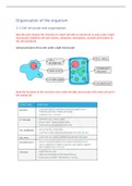
Describe and compare the structure of a plant cell with an animal cell, as seen under a light
microscope, limited to cell wall, nucleus, cytoplasm, chloroplasts, vacuoles and location of
the cell membrane
Animal and plant cell as seen under a light microscope:
State the functions of the structures seen under the light microscope in the plant cell and in
the animal cell
, State that almost all cells, except prokaryotes, have mitochondria, rough endoplasmic
reticulum, ribosomes on rough endoplasmic reticulum and vesicles
Within the cytoplasm, the following organelles are visible in almost all cells except
prokaryotes when looking at higher magnification (i.e. using an electron microscope):
Mitochondria: organelles found throughout the cytoplasm that are the site of
aerobic respiration
Ribosomes: site of protein synthesis. Can be free within the cytoplasm or attached to
a system of membranes within the cell known as Endoplasmic Reticulum (a network
of membranous tubules within the cytoplasm of a eukaryotic cell, continuous with
the nuclear membrane)
Endoplasmic reticulum studded with ribosomes looks rough under the microscope;
this gives rise to its name of Rough Endoplasmic Reticulum (often shortened
to R.E.R.)
Vesicles can also be seen using a higher magnification: these are small circular
structures found moving throughout the cytoplasm
Identify mitochondria and rough endoplasmic reticulum in diagrams and images of cells
State that cells with high rates of metabolism require large numbers of mitochondria to
provide sufficient energy
2.2 Levels of organisation
Specialised cells are those which have developed certain characteristics in order to perform
particular functions. These differences are controlled by genes in the nucleus (switching
genes on/off).
Cells specialise by undergoing differentiation: this is a process by which cells develop the
structure and characteristics needed to be able to carry out their functions.
Relate the structure of the following to their functions:
2.1 Cell structure and organisation
Describe and compare the structure of a plant cell with an animal cell, as seen under a light
microscope, limited to cell wall, nucleus, cytoplasm, chloroplasts, vacuoles and location of
the cell membrane
Animal and plant cell as seen under a light microscope:
State the functions of the structures seen under the light microscope in the plant cell and in
the animal cell
, State that almost all cells, except prokaryotes, have mitochondria, rough endoplasmic
reticulum, ribosomes on rough endoplasmic reticulum and vesicles
Within the cytoplasm, the following organelles are visible in almost all cells except
prokaryotes when looking at higher magnification (i.e. using an electron microscope):
Mitochondria: organelles found throughout the cytoplasm that are the site of
aerobic respiration
Ribosomes: site of protein synthesis. Can be free within the cytoplasm or attached to
a system of membranes within the cell known as Endoplasmic Reticulum (a network
of membranous tubules within the cytoplasm of a eukaryotic cell, continuous with
the nuclear membrane)
Endoplasmic reticulum studded with ribosomes looks rough under the microscope;
this gives rise to its name of Rough Endoplasmic Reticulum (often shortened
to R.E.R.)
Vesicles can also be seen using a higher magnification: these are small circular
structures found moving throughout the cytoplasm
Identify mitochondria and rough endoplasmic reticulum in diagrams and images of cells
State that cells with high rates of metabolism require large numbers of mitochondria to
provide sufficient energy
2.2 Levels of organisation
Specialised cells are those which have developed certain characteristics in order to perform
particular functions. These differences are controlled by genes in the nucleus (switching
genes on/off).
Cells specialise by undergoing differentiation: this is a process by which cells develop the
structure and characteristics needed to be able to carry out their functions.
Relate the structure of the following to their functions: