Structural biology: X-Ray Crystallography
The 3 pillars that structural biology is built on are NMR, ETM & X-Ray Crystallography.
Why use protein crystallography?
• Gives a lot of definitive structural information
• Good for studying tight complexes (inhibitors, drugs)
• Relatively fast (once gotten crystal
- it takes 20 min to solve structure from crystal
- NMR takes 18 months
Limitations of protein crystallography?
• Need pure protein in mg quantities
• Need to grow crystals that diffract
• Get a time and space average
When to use NMR?
• Investigating protein folding and dynamics
• Studying loosely associating molecules
• Only other technique available if can’t grow crystal
Limitations of NMR
• Proteins must be le than 30,000 Daltons
• Proteins must be non-aggregating at high concentrations (as require high concentration in the
NMR tube) and low Ph.
• Relatively slow (in obtaining data) compared to X-ray methods
Other techniques
• Transmission electron microscopy (TEM)
• Atomical force microscopy (AFM)
Overview:
1. Protein crystals
2. X-ray diffraction
3. The unit cell and crystal lattice
4. Braggs law and resolution
5. Symmetry and space groups
6. Data processing and scaling
7. The phase problem
8. Phasing methods
,Introduction
• X-ray crystallography uses X-rays and crystals.
• X-rays interact weakly with matter
• In X-ray crystallography, use Angstrom as unit
• Angstrom is not an SI unit
• Angstrom id 10-10 M
• The C-C bond length is 1.5A
X-Ray crystallography
Why crystals?
• Crystals arrange molecules in a precise way in 3D space to get a strong diffraction pattern
• The crystal is a source of amplifier of the signal because a crystal has many molecules arranged
in a regular way and amplifies the diffraction pattern achieved
Why X-rays?
• We use X-rays because they have the right wavelength that want to resolve, which 1.5A C-C
bond length is.
• Crystallographers deposit there protein structure in a protein data bank
Macromolecular crystallography
• Macromolecular crystallography refers to protein or nucleic acids
• Crystallographers deposit their proteins structure in the protein data bank
• Spots are Braggs deflection
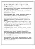
,Examples of crystals
•
Crystals from inorganic and especially from ionic
compounds like NaCl
• Crystals are stable.
- kept at room temperature for long time in dry
environment
• Sometimes proteins are crystallised by using
ammonium sulphate solution
• How to tell if have protein or salt crystal
- Protein crystals are v.fragile compared to salt
crystals.
• Ice, salt and sugar are examples of everyday crystals
• Salt crystals are dry with little water
Protein crystals
• Proteins are generally more sensitive than mineral
- Grown from solution and kept in humid environment throughout experiment
• Protein crystals are 50% water and 50% protein
• Protein crystals require water to give a diffraction pattern
• Protein crystals grown size 0.1 to 1mm.
• At high temperature, protein degenerate
What are X-rays?
• X-rays and visible light are part of the electromagnetic spectrum
• Visible light is composed of electromagnetic waves
- Every colour has a wavelength energy
• X-rays are the same but with higher energy i.e. shorter wavelength
• To distinguish objects, which are a distant d apart use light with wavelength λ ≤ 2d.
• C-C bond length is about 1.5Å so light with λ ≤ 3Å required: X-rays
• The wavelength of light is 400 nm = 4000Angstrom
• The smallest thing can see is in the range of 400nm
• If use visible wavelength of light will not be able to see the protein let alone the C-C bonds
- If want to see anatomical structure need to use wavelength of X-ray
• Therefore, we use X-rays because they have the right wavelength
• Monochromatic=single wavelength
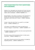
, Principle of an X-ray experiment
• Crystal diffracts in all directions.
• The detector cannot cover the full sphere around the crystal, crystal is rotated during
experiment
• 1 typical data set consists of tens of hundreds of images and the crystal is rotated for between90
and 360 degrees.
• Position of spots= how molecules are packed in the crystal
• Spot intensity= molecular structure information
• 0.2mm3 is the volume of the crystal
• Rotate the crystal 90, 180, 360 degrees depending on the symmetry of the crystal
• The higher the symmetry of the crystal, the fewer degrees of rotation have to go through
The 3 pillars that structural biology is built on are NMR, ETM & X-Ray Crystallography.
Why use protein crystallography?
• Gives a lot of definitive structural information
• Good for studying tight complexes (inhibitors, drugs)
• Relatively fast (once gotten crystal
- it takes 20 min to solve structure from crystal
- NMR takes 18 months
Limitations of protein crystallography?
• Need pure protein in mg quantities
• Need to grow crystals that diffract
• Get a time and space average
When to use NMR?
• Investigating protein folding and dynamics
• Studying loosely associating molecules
• Only other technique available if can’t grow crystal
Limitations of NMR
• Proteins must be le than 30,000 Daltons
• Proteins must be non-aggregating at high concentrations (as require high concentration in the
NMR tube) and low Ph.
• Relatively slow (in obtaining data) compared to X-ray methods
Other techniques
• Transmission electron microscopy (TEM)
• Atomical force microscopy (AFM)
Overview:
1. Protein crystals
2. X-ray diffraction
3. The unit cell and crystal lattice
4. Braggs law and resolution
5. Symmetry and space groups
6. Data processing and scaling
7. The phase problem
8. Phasing methods
,Introduction
• X-ray crystallography uses X-rays and crystals.
• X-rays interact weakly with matter
• In X-ray crystallography, use Angstrom as unit
• Angstrom is not an SI unit
• Angstrom id 10-10 M
• The C-C bond length is 1.5A
X-Ray crystallography
Why crystals?
• Crystals arrange molecules in a precise way in 3D space to get a strong diffraction pattern
• The crystal is a source of amplifier of the signal because a crystal has many molecules arranged
in a regular way and amplifies the diffraction pattern achieved
Why X-rays?
• We use X-rays because they have the right wavelength that want to resolve, which 1.5A C-C
bond length is.
• Crystallographers deposit there protein structure in a protein data bank
Macromolecular crystallography
• Macromolecular crystallography refers to protein or nucleic acids
• Crystallographers deposit their proteins structure in the protein data bank
• Spots are Braggs deflection
,Examples of crystals
•
Crystals from inorganic and especially from ionic
compounds like NaCl
• Crystals are stable.
- kept at room temperature for long time in dry
environment
• Sometimes proteins are crystallised by using
ammonium sulphate solution
• How to tell if have protein or salt crystal
- Protein crystals are v.fragile compared to salt
crystals.
• Ice, salt and sugar are examples of everyday crystals
• Salt crystals are dry with little water
Protein crystals
• Proteins are generally more sensitive than mineral
- Grown from solution and kept in humid environment throughout experiment
• Protein crystals are 50% water and 50% protein
• Protein crystals require water to give a diffraction pattern
• Protein crystals grown size 0.1 to 1mm.
• At high temperature, protein degenerate
What are X-rays?
• X-rays and visible light are part of the electromagnetic spectrum
• Visible light is composed of electromagnetic waves
- Every colour has a wavelength energy
• X-rays are the same but with higher energy i.e. shorter wavelength
• To distinguish objects, which are a distant d apart use light with wavelength λ ≤ 2d.
• C-C bond length is about 1.5Å so light with λ ≤ 3Å required: X-rays
• The wavelength of light is 400 nm = 4000Angstrom
• The smallest thing can see is in the range of 400nm
• If use visible wavelength of light will not be able to see the protein let alone the C-C bonds
- If want to see anatomical structure need to use wavelength of X-ray
• Therefore, we use X-rays because they have the right wavelength
• Monochromatic=single wavelength
, Principle of an X-ray experiment
• Crystal diffracts in all directions.
• The detector cannot cover the full sphere around the crystal, crystal is rotated during
experiment
• 1 typical data set consists of tens of hundreds of images and the crystal is rotated for between90
and 360 degrees.
• Position of spots= how molecules are packed in the crystal
• Spot intensity= molecular structure information
• 0.2mm3 is the volume of the crystal
• Rotate the crystal 90, 180, 360 degrees depending on the symmetry of the crystal
• The higher the symmetry of the crystal, the fewer degrees of rotation have to go through