vEC leerdoelen periode 5
Anatomie van de lever
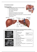
De ligging en (topografische) anatomie van de lever beschrijven:
o Linker leverkwab (lobus sinister)
o Rechter leverkwab (lobus dexter)
o Lobus caudatus → ligt craniaal
o Lobus quadratus
o Ligamentum teres (afgrenzing van lobus quadratus)
o Interlobaire fissuur (afgrenzing van lobus caudatus)
o Ligamentum falciforme (niet te zien op de echo → in anatomie de scheidingslijn
tussen dextra en sinistra)
o Leverpoort/hilus;
Op het echografische beeld van de lever de structuren benoemen en aanwijzen;
1. Vena portae ramus sinistra
2. Hart
3. Vena hepatica media
4. Vena cava inferior Sagittaal
5. Confluens Linker leverkwab
6. Arteria hepatica communis
7. Lobus caudatus
8. Interlobaire fissuur
1. Rectus abdominalis/buikspier
2. Ligamentum teres
Transversaal
3. Venae porta ramus sinistra
4. Vena cava inferior
5. Vena hepatica media
6. (Interlobaire fissuur)/slagschaduw van ligamentum teres
7. Pancreas cauda
8. Aorta abdominalis
, 1. Caudale leverpunt
2. Diafragma
3. Rechter nier
4. Rechter leverkwab Sagittaal
1. Vena porta takje
2. Vena hepatica dextra
3. Vena hepatica dextra
4. Rechter leverkwab Transversaal
5. Vena hepatica media
6. Vena cava inferior
7. Diafragma
1. galblaas
2. vena portae intrahepatisch
3. rechter leverkwab
4. ligamentum teres Oblique
5. lobus quadratus
6. vena cava inferior
1. Latero-ventrale leverrand
Oblique
2. Diafragma (lateraal)
Vena hepatica dextra
3. Vena portae ramus dextra
4. Spiegelartefact (niet duidelijk,
wel plaats waar vaak zichtbaar)/lon
Aan de hand van een echografische opname beschrijven in welke richting de genummerde
structuren/ vaten zijn afgebeeld.
Verschillende systemen in de lever benoemen
o Portale (bloedvaten) systeem
Vena porta voert bloed af met
afvalstoffen de lever in
o Veneuze systeem
Loopt af naar craniaal, voeren
af naar de vci
o Arteriële systeem
Voert zuurstofrijk bloed aan
o Galwegen
Stroom de lever uit, gal moet
naar de darmen
Anatomie van de lever
De ligging en (topografische) anatomie van de lever beschrijven:
o Linker leverkwab (lobus sinister)
o Rechter leverkwab (lobus dexter)
o Lobus caudatus → ligt craniaal
o Lobus quadratus
o Ligamentum teres (afgrenzing van lobus quadratus)
o Interlobaire fissuur (afgrenzing van lobus caudatus)
o Ligamentum falciforme (niet te zien op de echo → in anatomie de scheidingslijn
tussen dextra en sinistra)
o Leverpoort/hilus;
Op het echografische beeld van de lever de structuren benoemen en aanwijzen;
1. Vena portae ramus sinistra
2. Hart
3. Vena hepatica media
4. Vena cava inferior Sagittaal
5. Confluens Linker leverkwab
6. Arteria hepatica communis
7. Lobus caudatus
8. Interlobaire fissuur
1. Rectus abdominalis/buikspier
2. Ligamentum teres
Transversaal
3. Venae porta ramus sinistra
4. Vena cava inferior
5. Vena hepatica media
6. (Interlobaire fissuur)/slagschaduw van ligamentum teres
7. Pancreas cauda
8. Aorta abdominalis
, 1. Caudale leverpunt
2. Diafragma
3. Rechter nier
4. Rechter leverkwab Sagittaal
1. Vena porta takje
2. Vena hepatica dextra
3. Vena hepatica dextra
4. Rechter leverkwab Transversaal
5. Vena hepatica media
6. Vena cava inferior
7. Diafragma
1. galblaas
2. vena portae intrahepatisch
3. rechter leverkwab
4. ligamentum teres Oblique
5. lobus quadratus
6. vena cava inferior
1. Latero-ventrale leverrand
Oblique
2. Diafragma (lateraal)
Vena hepatica dextra
3. Vena portae ramus dextra
4. Spiegelartefact (niet duidelijk,
wel plaats waar vaak zichtbaar)/lon
Aan de hand van een echografische opname beschrijven in welke richting de genummerde
structuren/ vaten zijn afgebeeld.
Verschillende systemen in de lever benoemen
o Portale (bloedvaten) systeem
Vena porta voert bloed af met
afvalstoffen de lever in
o Veneuze systeem
Loopt af naar craniaal, voeren
af naar de vci
o Arteriële systeem
Voert zuurstofrijk bloed aan
o Galwegen
Stroom de lever uit, gal moet
naar de darmen