

The Nervous System: A Communication Network
Behaviour depends on rapid information travel and processing. The nervous system is the
body’s communication network, handling information just as the circulatory system handles
blood. The basic components of the nervous system are two types of living cells called neurons
and glia.
Neurons are cells that receive, integrate, and transmit information. Their function is
communication. They are specialized cells that carry information throughout the nervous system.
Glia are cells that provide structural support, insulate, and communicate. Their function is to
act as neural glue and provide nourishment to the neuron.
Neurons
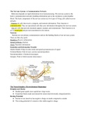
Neurons are our primary communicators and are the building blocks of our nervous system.
There are three key parts:
Dendrites: Receive information
Soma/Cell Body: Nucleus
Axon: Transmits information
Insolation and Information Transfer
Myelin Sheath: Wrap on some axons and speed up transmission of signal
Terminal Button: End of axon; secretes neurotransmitters
Neurotransmitters: Chemical messengers
Synapse: Point at which neurons interconnect
The Neural Impulse: Electrochemical Beginnings
Hodgkin and Huxley
● Studied giant squids since squids have large axons
● Found that fluids inside and outside the neuron had electrically charged particles
Resting Potential
● Neuron at rest (inactive) has negative charge on inside compared to outside
● The resting potential of a neuron is this stable negative charge
, Action Potential
● Stimulation causes the cell membrane to open briefly
● Positively charged sodium ions from outside the cell flow in
● Shift in electrical charge travels along neuron’s axon
● Action potential occurs
● Action potential functions according to an “All-or-None Law”
The Synapse: Where Neurons Meet
Synaptic Cleft: The ‘space’ between terminal button of one neuron and cell membrane of
another. Electrical signals cannot jump the gap so they need the help of neurotransmitters.
Presynaptic Neuron: Sends messages via synaptic vesicles (sacs that release neurotransmitters).
The NTs are released into the cleft.
Postsynaptic Neuron: Receives messages via its reception sites.
When a neural impulse reaches an axon’s terminal buttons, it triggers the release of
chemical messengers called neurotransmitters.The neurotransmitter molecules diffuse across the
synaptic cleft and bind to receptor sites on the postsynaptic neuron.A specific neurotransmitter
can bind only to receptor sites that its molecular structure will fit into, much like a key must fit a
lock.
Transmission
1. Synthesis - Neurotransmitters manufactured by cell body
2. Transportation and Storage - synaptic vessels
3. Release - presynaptic, neurotransmitters send to synaptic cleft
4. Binding - receptors
5. Deactivation
6. Autoreceptor Activation
7. Reuptake
8. Degradation
Receiving Signals: Pre and Postsynaptic Potentials
A voltage change at the receptor site is called the postsynaptic potential. It is not all-or-none
but it changes the probability of the postsynaptic neuron firing. A positive voltage shift results in
excitatory postsynaptic potential while a negative voltage shift results in inhibitory postsynaptic
potential.
Integrating Signals, Neural Networks, and Synaptic Connections
One neuron receives signals from thousands of other neurons. This requires integration of
signals. So, PSPs add up and balance out. Once there is balance between IPSPs and EPSPs there
is graded potential. Neural networks are interconnected neurons that fire together sequentially.
Specific neurotransmitters work at specific synapses. This is like a lock and key mechanism;
not all neurotransmitters fit into the postsynaptic cell. The agonist will mimic the action of a
Behaviour depends on rapid information travel and processing. The nervous system is the
body’s communication network, handling information just as the circulatory system handles
blood. The basic components of the nervous system are two types of living cells called neurons
and glia.
Neurons are cells that receive, integrate, and transmit information. Their function is
communication. They are specialized cells that carry information throughout the nervous system.
Glia are cells that provide structural support, insulate, and communicate. Their function is to
act as neural glue and provide nourishment to the neuron.
Neurons
Neurons are our primary communicators and are the building blocks of our nervous system.
There are three key parts:
Dendrites: Receive information
Soma/Cell Body: Nucleus
Axon: Transmits information
Insolation and Information Transfer
Myelin Sheath: Wrap on some axons and speed up transmission of signal
Terminal Button: End of axon; secretes neurotransmitters
Neurotransmitters: Chemical messengers
Synapse: Point at which neurons interconnect
The Neural Impulse: Electrochemical Beginnings
Hodgkin and Huxley
● Studied giant squids since squids have large axons
● Found that fluids inside and outside the neuron had electrically charged particles
Resting Potential
● Neuron at rest (inactive) has negative charge on inside compared to outside
● The resting potential of a neuron is this stable negative charge
, Action Potential
● Stimulation causes the cell membrane to open briefly
● Positively charged sodium ions from outside the cell flow in
● Shift in electrical charge travels along neuron’s axon
● Action potential occurs
● Action potential functions according to an “All-or-None Law”
The Synapse: Where Neurons Meet
Synaptic Cleft: The ‘space’ between terminal button of one neuron and cell membrane of
another. Electrical signals cannot jump the gap so they need the help of neurotransmitters.
Presynaptic Neuron: Sends messages via synaptic vesicles (sacs that release neurotransmitters).
The NTs are released into the cleft.
Postsynaptic Neuron: Receives messages via its reception sites.
When a neural impulse reaches an axon’s terminal buttons, it triggers the release of
chemical messengers called neurotransmitters.The neurotransmitter molecules diffuse across the
synaptic cleft and bind to receptor sites on the postsynaptic neuron.A specific neurotransmitter
can bind only to receptor sites that its molecular structure will fit into, much like a key must fit a
lock.
Transmission
1. Synthesis - Neurotransmitters manufactured by cell body
2. Transportation and Storage - synaptic vessels
3. Release - presynaptic, neurotransmitters send to synaptic cleft
4. Binding - receptors
5. Deactivation
6. Autoreceptor Activation
7. Reuptake
8. Degradation
Receiving Signals: Pre and Postsynaptic Potentials
A voltage change at the receptor site is called the postsynaptic potential. It is not all-or-none
but it changes the probability of the postsynaptic neuron firing. A positive voltage shift results in
excitatory postsynaptic potential while a negative voltage shift results in inhibitory postsynaptic
potential.
Integrating Signals, Neural Networks, and Synaptic Connections
One neuron receives signals from thousands of other neurons. This requires integration of
signals. So, PSPs add up and balance out. Once there is balance between IPSPs and EPSPs there
is graded potential. Neural networks are interconnected neurons that fire together sequentially.
Specific neurotransmitters work at specific synapses. This is like a lock and key mechanism;
not all neurotransmitters fit into the postsynaptic cell. The agonist will mimic the action of a


