Inductive reasoning - From specific knowledge to general knowledge. IN MOST CASES,
FOR BIO AND CHEM, ITS BETTER TO USE INDUCTIVE REASONING!
Deductive reasoning - From general knowledge to specific reasoning.
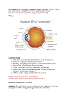
The eye
Function of life
1. Metabolism - chemical reaction that release energy for cellular use
2. Response - respond to internal/external stimuli
3. Sensitivity - living things are responsive to internal and external stimuli
4. Homeostasis - maintain stable internal conditions
5. Growth - increase in cell size or cell number, grow/develop
6. Reproduction - produce more offspring/cells - sexually or asexually
7. Excretion - remove waste products
8. Nutrition - obtain energy / materials from environment
Response = action to internal or external stimuli
Sensitivity = the ability to detect internal or external stimuli
Metabolism = Anabolism + catabolism
Anabolism - the synthesis of complex molecules in living organisms from simpler
ones together with the storage of energy. Constructive metabolism
,Catabolism - the breaking down of complex molecules in living organisms to form
simpler ones, together with the release of energy. Destructive metabolism
Cell theory states that:
1. All living things are made up of cells
2. Cells are the basic unit and structure of life
3. Cells arise from pre-existing cells
Prokaryote’s cell wall is made of peptidoglycan
Eukaryote’s cell wall is made up of:
- Plants -> cellulose
- Fungi -> Chitin
Dyes you can use to stain microorganisms:
1. Methylene blue
2. Crystal violet
3. Safranin (counter stain)
- Usually use ethanol or asanol
Bacteria can be classified by the reaction to the dye:
1. Gram positive bacteria appear blue
- The peptidoglycan layer in gram positive bacteria is thicker than gram
negative.
- Gram positive bacteria easily absorb stains.
- Antibiotics weakens the cell wall / the peptidoglycan layer, making it easier for
the bacteria to die.
2. Gram negative bacteria appears red or pink
- Gram positive has two outer layers, so it
- Since the peptidoglycan layer is thinner, it is harder to absorb. Not only that,
but the outer membrane blocks a bit of the stain, making it harder for stains to
see.
- The outer layer is used to somewhat block antibiotics, so antibodies have a
harder time killing gram negative bacteria.
Pili / pilus of a prokaryote is used to attach to skin surfaces.
Svedberg unit - used to find the sedimentation rate
Small subunit (30s) + large subunit (50s) = complete 70s Ribosome
Nucleolus - Part of a nucleus that creates ribosomes
Proteins absorb dyes, so when there is a high amount of protein, it will look very dark under
an electron microscope.
Nucleoid - A region that contains genetic material / DNA
Cell organelle Prokaryote Eukaryote
Ribosome Size: 70S Size: 80S
,Coarse adjustment nob is used to move the stage up and down / control the elevation of the
stage.
FIne adjustment nob is used to control the clarity / focus of the lens to view the specimen
clearly.
The diaphragm is used to control the light intensity / amount of light the (Illuminator / light
source)
Compound microscopes uses multiple lenses, one in the eye piece, and the other as an
objective lens. They tend to have limitations in clarity and magnification.
In more advanced laboratories, they use SEM and TEM for a clearer view of the specimen:
1. Scanning ELectron Microscope, uses the beams of electrons. Electrons will be fired
to the specimen and reflected back to the eye piece, giving a clearer image. This is
used to focus on the sample’s surface and its compositions.
2. Transmission Electron Microscope, this is used to provide details of the internal
composition of the sample.
, 3. Fluorescence microscope, it can view any images that are actually fluorescent
stained, meaning that the specimen is treated with stains. Stains are actually
proteins. Heavily stained parts tend to be the nucleus. Under the microscope, they
will be glowing. It has a greater resolution that SEM and TEM. Immunofluorescence
When using a microscope, the sample will appear black and white. The
Plasmid
WHAT ARE PLASMIDS?
- A plasmid is a small, circular piece of DNA that is different than the
chromosomal DNA, which is all the genetic material found in an organism’s
chromosomes. It replicates independently of chromosomal DNA.
- Mainly found in Bacteria, but also can be found in archaea and multicellular
organisms.
- Usually carry 1 gene which is beneficial to their host organisms.
- Not considered to be an independent life.
- Replication of a genetic material is formed then transferred to the other
bacterias. (so that other bacterias can be resistant)
- THe single chromosome in a bacteria is used for life functions
- Plasmids are used for survival (from environmental stressors)
- Plasmids can join together with the single chromosome to get more chances
of survival.
- Traits can be passed down from one bacteria to another
Types of plasmids:
1. Antibiotic resistance = resistance plasmid
2. Degradative plasmid = a metabolic plasmid which breaks down waste
(biodegradation)
3. Catabolic plasmid =
4. Virulence plasmid = Plasmids that can cause an infection
5. Toxin plasmid = Plasmids that produces toxins(antigens / proteins)
6. Fertility plasmid = transfers plasmids to another prokaryotic cell and replicates
plasmids
7. Heat-resistant plasmid
Virulence = danger level
Bacterial conjugation - the transfer of genetic material between bacterial cells by
direct cell-to-cell contact or by a bridge-like connection between two cells. Simple
definition, a pili will start to stretch and when two pilis connect, a transfer of genetic
material occurs.
FOR BIO AND CHEM, ITS BETTER TO USE INDUCTIVE REASONING!
Deductive reasoning - From general knowledge to specific reasoning.
The eye
Function of life
1. Metabolism - chemical reaction that release energy for cellular use
2. Response - respond to internal/external stimuli
3. Sensitivity - living things are responsive to internal and external stimuli
4. Homeostasis - maintain stable internal conditions
5. Growth - increase in cell size or cell number, grow/develop
6. Reproduction - produce more offspring/cells - sexually or asexually
7. Excretion - remove waste products
8. Nutrition - obtain energy / materials from environment
Response = action to internal or external stimuli
Sensitivity = the ability to detect internal or external stimuli
Metabolism = Anabolism + catabolism
Anabolism - the synthesis of complex molecules in living organisms from simpler
ones together with the storage of energy. Constructive metabolism
,Catabolism - the breaking down of complex molecules in living organisms to form
simpler ones, together with the release of energy. Destructive metabolism
Cell theory states that:
1. All living things are made up of cells
2. Cells are the basic unit and structure of life
3. Cells arise from pre-existing cells
Prokaryote’s cell wall is made of peptidoglycan
Eukaryote’s cell wall is made up of:
- Plants -> cellulose
- Fungi -> Chitin
Dyes you can use to stain microorganisms:
1. Methylene blue
2. Crystal violet
3. Safranin (counter stain)
- Usually use ethanol or asanol
Bacteria can be classified by the reaction to the dye:
1. Gram positive bacteria appear blue
- The peptidoglycan layer in gram positive bacteria is thicker than gram
negative.
- Gram positive bacteria easily absorb stains.
- Antibiotics weakens the cell wall / the peptidoglycan layer, making it easier for
the bacteria to die.
2. Gram negative bacteria appears red or pink
- Gram positive has two outer layers, so it
- Since the peptidoglycan layer is thinner, it is harder to absorb. Not only that,
but the outer membrane blocks a bit of the stain, making it harder for stains to
see.
- The outer layer is used to somewhat block antibiotics, so antibodies have a
harder time killing gram negative bacteria.
Pili / pilus of a prokaryote is used to attach to skin surfaces.
Svedberg unit - used to find the sedimentation rate
Small subunit (30s) + large subunit (50s) = complete 70s Ribosome
Nucleolus - Part of a nucleus that creates ribosomes
Proteins absorb dyes, so when there is a high amount of protein, it will look very dark under
an electron microscope.
Nucleoid - A region that contains genetic material / DNA
Cell organelle Prokaryote Eukaryote
Ribosome Size: 70S Size: 80S
,Coarse adjustment nob is used to move the stage up and down / control the elevation of the
stage.
FIne adjustment nob is used to control the clarity / focus of the lens to view the specimen
clearly.
The diaphragm is used to control the light intensity / amount of light the (Illuminator / light
source)
Compound microscopes uses multiple lenses, one in the eye piece, and the other as an
objective lens. They tend to have limitations in clarity and magnification.
In more advanced laboratories, they use SEM and TEM for a clearer view of the specimen:
1. Scanning ELectron Microscope, uses the beams of electrons. Electrons will be fired
to the specimen and reflected back to the eye piece, giving a clearer image. This is
used to focus on the sample’s surface and its compositions.
2. Transmission Electron Microscope, this is used to provide details of the internal
composition of the sample.
, 3. Fluorescence microscope, it can view any images that are actually fluorescent
stained, meaning that the specimen is treated with stains. Stains are actually
proteins. Heavily stained parts tend to be the nucleus. Under the microscope, they
will be glowing. It has a greater resolution that SEM and TEM. Immunofluorescence
When using a microscope, the sample will appear black and white. The
Plasmid
WHAT ARE PLASMIDS?
- A plasmid is a small, circular piece of DNA that is different than the
chromosomal DNA, which is all the genetic material found in an organism’s
chromosomes. It replicates independently of chromosomal DNA.
- Mainly found in Bacteria, but also can be found in archaea and multicellular
organisms.
- Usually carry 1 gene which is beneficial to their host organisms.
- Not considered to be an independent life.
- Replication of a genetic material is formed then transferred to the other
bacterias. (so that other bacterias can be resistant)
- THe single chromosome in a bacteria is used for life functions
- Plasmids are used for survival (from environmental stressors)
- Plasmids can join together with the single chromosome to get more chances
of survival.
- Traits can be passed down from one bacteria to another
Types of plasmids:
1. Antibiotic resistance = resistance plasmid
2. Degradative plasmid = a metabolic plasmid which breaks down waste
(biodegradation)
3. Catabolic plasmid =
4. Virulence plasmid = Plasmids that can cause an infection
5. Toxin plasmid = Plasmids that produces toxins(antigens / proteins)
6. Fertility plasmid = transfers plasmids to another prokaryotic cell and replicates
plasmids
7. Heat-resistant plasmid
Virulence = danger level
Bacterial conjugation - the transfer of genetic material between bacterial cells by
direct cell-to-cell contact or by a bridge-like connection between two cells. Simple
definition, a pili will start to stretch and when two pilis connect, a transfer of genetic
material occurs.