Whole summary hap
Lecture 1 introduction to the heart
- Cardiomyocyte: muscle cell contracting in the heart
- Depolarization refers to the movement of a cell's membrane
potential to a more positive value
- repolarization refers to the change in membrane potential,
returning to a negative value
Two circulations:
1. Pulmonary circulation: goes to the lungs
2. Systemic circulation: goes to the rest of the body
Difference between the two:
o Difference in concentration of oxygen in the blood
o Pressure
▪ Pulmonary: low pressure
▪ Systemic: high pressure
- No separation between the circulations→ always low pressure
Heart
Function of the heart
- Pumping deoxygenated blood to the lungs
- Pumping oxygenated blood to all the organs in the body
- Together with blood vessels: providing adequate perfusion of all organs & tissues of the body
- Contraction and relaxation determine cardiac output
o Contraction: blood going out of the heart
o Relaxation: blood going into the heart
- The heartbeat is coordinated by contraction and relaxation of 2-3 billion CMs
Excitation-contraction coupling
- Contraction of the heart following electrical stimulation of cardiomyocytes
Automation of the heart
- The heart can beat independent of hormonal or neuronal input
- When the heart is outside the body the heart will still beat
o Because of spontaneous active pacemaker cells
Conduction through the heart
SA node:
- present in the right atrium
- contains pacemaker cells
o starts heartbeat
Conduction between cardiomyocytes
- The signal spreads through the neuronal cells with bundle branches
,Ion channels & action potential of ventricular cell
Membrane potential: determined by concentrations
differences of ions and permeability to ions
- Largely determined by K+ gradient
- Na: higher concentration outside of the cell,
channel open just after the peak
- Ca: higher concentration outside of the cell,
channel open just before peak
- K: higher concentration in the cell, channel
opens later, cell becomes more negative
Adrenaline changes the heartrate before K entrance
- Unstable resting potential - Stabile resting potential: –85 mV
- Slow depolarisation - Quick depolarisation
prepotential(pacemaker potential) - Plateau
Heart rate
- Determined by:
o Resting membrane potential of SA node cells
o Velocity of depolarisation: slope of the prepotential
Heart rate during exercise
- Maintain perfusion in times of increased demand
o Blood flow increases: heart rate increases
▪ Skeletal muscles increase the most in ml/min
Sympathetic stimulation
- Caused by noradrenaline/epinephrine
- Opens Ca2+ & Na+ channels
- Quicker depolarisation = Steeper pacemaker potent
- Less negative resting potential
- Active in activity
Parasympathetic stimulation
- Caused by acetylcholine
- Opens K+ channels
- Active in rest
,Refractory period
- Period in which cells are inexcitable (Na+-channels are not reset)
- Absolute and relative refractory periods
- Key to contraction relaxation behaviour of cardiomyocytes
What if something goes wrong with the action potential?
- Mutation in 1 of the ion channels, causing impaired repolarisation:
o Long QT syndrome ->
Ca2+ and contraction
- C.I.C.R. = calcium induced calcium release
- Ca2+-binding to myofilament initiates contraction
- Myosin binds to actine in movement
- Rest: proteins blocking actine
- No ATP: Ca stays high -> stays contracted
o ATP: breaks myosin from actine
Development of cardiomyocytes forced by:
- Amount of intracytosolair calcium
- Ca2+-sensitivity of contractile apparatus
A single heart beat at cellular level
- Electrical signal from neighbouring cell (CM, SA node, conduction system)
- Action potential (1. Na+ influx; 2. Ca2+ influx; 3. K+ efflux)
- Ca2+ induced Ca2+ release
- Ca2+ binding to myofilaments
- Power stroke => cell shortening
Ca2+-release from myofilamentsReuptake in SR => relaxation
Ventricle cell
- Sodium entering
- More important: Ca entering,
o High ca concentration, contraction
- Ca cycling
o Needed for big cells
- SERCA: organelles in muscles filled with Ca
o Released when Ca is present in the cells
o 10 fold increase in Ca
Action potential & ECG
Linked but separate events!
What is measured in an ECG
- Individual action potentials are not measured
- ECG signal reflects electrical differences
o White muscle/signal: rest, no signal
o Red muscle/signal: active signal
Signal of ECG
- determined by:
, o location of electrodes (how do you “look” at the heart)
o Distance of electrodes to the heart
o Size of the heart muscle (mass = size of depolarisation wave)
ECG of Einthoven
- Problem: Electrical activity could not be measured in patients
- Solution: developed the string galvanometer (thin conducting wire between 2 strong magnets).
Current running through the wire, electrical, movement of the wire →ECG
- Machine was too big for the hospital
o Telephone wire between the hospital and the lab
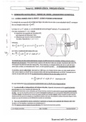
Einthovens triangle
- Convention: depolarisation wave towards positive electrode: Positive signal on ECG
- Lead I: horizontally right arm negative, left arm posi
- Lead II: right arm to left leg leg posi
- Lead III: left arm to left leg
Shape of the ECG
- + charge towards + electrode: positive peak
- + charge towards – electrode: negative peak
- - charge towards – electrode: positive peak
- P-wave:
o Atrial contraction/depolarisation
o Positive depolarisation charge moves towards the positive electrode and AV node
- PR interval:
o Electric charge stays in the AV node for a while
o AV node is depolarised but no movement
- Q-wave:
o Charge moves through the His bundle -> septal depolarisation
o Moves in the opposite direction from the positive electrode
o not always detected
- R-wave
o Charge moves to the edge of the heart
o Left ventricle has a larger charge
- S-wave:
o Charge moves upwards toward heart base
- QRS: Ventricular contraction
- ST segment:
o Ventricular relaxation
o Depolarisation of the ventricular myocardium
- T-wave:
o Ventricular repolarisation
o Positive charge becomes negative towards negative electrode
ECG
Lecture 1 introduction to the heart
- Cardiomyocyte: muscle cell contracting in the heart
- Depolarization refers to the movement of a cell's membrane
potential to a more positive value
- repolarization refers to the change in membrane potential,
returning to a negative value
Two circulations:
1. Pulmonary circulation: goes to the lungs
2. Systemic circulation: goes to the rest of the body
Difference between the two:
o Difference in concentration of oxygen in the blood
o Pressure
▪ Pulmonary: low pressure
▪ Systemic: high pressure
- No separation between the circulations→ always low pressure
Heart
Function of the heart
- Pumping deoxygenated blood to the lungs
- Pumping oxygenated blood to all the organs in the body
- Together with blood vessels: providing adequate perfusion of all organs & tissues of the body
- Contraction and relaxation determine cardiac output
o Contraction: blood going out of the heart
o Relaxation: blood going into the heart
- The heartbeat is coordinated by contraction and relaxation of 2-3 billion CMs
Excitation-contraction coupling
- Contraction of the heart following electrical stimulation of cardiomyocytes
Automation of the heart
- The heart can beat independent of hormonal or neuronal input
- When the heart is outside the body the heart will still beat
o Because of spontaneous active pacemaker cells
Conduction through the heart
SA node:
- present in the right atrium
- contains pacemaker cells
o starts heartbeat
Conduction between cardiomyocytes
- The signal spreads through the neuronal cells with bundle branches
,Ion channels & action potential of ventricular cell
Membrane potential: determined by concentrations
differences of ions and permeability to ions
- Largely determined by K+ gradient
- Na: higher concentration outside of the cell,
channel open just after the peak
- Ca: higher concentration outside of the cell,
channel open just before peak
- K: higher concentration in the cell, channel
opens later, cell becomes more negative
Adrenaline changes the heartrate before K entrance
- Unstable resting potential - Stabile resting potential: –85 mV
- Slow depolarisation - Quick depolarisation
prepotential(pacemaker potential) - Plateau
Heart rate
- Determined by:
o Resting membrane potential of SA node cells
o Velocity of depolarisation: slope of the prepotential
Heart rate during exercise
- Maintain perfusion in times of increased demand
o Blood flow increases: heart rate increases
▪ Skeletal muscles increase the most in ml/min
Sympathetic stimulation
- Caused by noradrenaline/epinephrine
- Opens Ca2+ & Na+ channels
- Quicker depolarisation = Steeper pacemaker potent
- Less negative resting potential
- Active in activity
Parasympathetic stimulation
- Caused by acetylcholine
- Opens K+ channels
- Active in rest
,Refractory period
- Period in which cells are inexcitable (Na+-channels are not reset)
- Absolute and relative refractory periods
- Key to contraction relaxation behaviour of cardiomyocytes
What if something goes wrong with the action potential?
- Mutation in 1 of the ion channels, causing impaired repolarisation:
o Long QT syndrome ->
Ca2+ and contraction
- C.I.C.R. = calcium induced calcium release
- Ca2+-binding to myofilament initiates contraction
- Myosin binds to actine in movement
- Rest: proteins blocking actine
- No ATP: Ca stays high -> stays contracted
o ATP: breaks myosin from actine
Development of cardiomyocytes forced by:
- Amount of intracytosolair calcium
- Ca2+-sensitivity of contractile apparatus
A single heart beat at cellular level
- Electrical signal from neighbouring cell (CM, SA node, conduction system)
- Action potential (1. Na+ influx; 2. Ca2+ influx; 3. K+ efflux)
- Ca2+ induced Ca2+ release
- Ca2+ binding to myofilaments
- Power stroke => cell shortening
Ca2+-release from myofilamentsReuptake in SR => relaxation
Ventricle cell
- Sodium entering
- More important: Ca entering,
o High ca concentration, contraction
- Ca cycling
o Needed for big cells
- SERCA: organelles in muscles filled with Ca
o Released when Ca is present in the cells
o 10 fold increase in Ca
Action potential & ECG
Linked but separate events!
What is measured in an ECG
- Individual action potentials are not measured
- ECG signal reflects electrical differences
o White muscle/signal: rest, no signal
o Red muscle/signal: active signal
Signal of ECG
- determined by:
, o location of electrodes (how do you “look” at the heart)
o Distance of electrodes to the heart
o Size of the heart muscle (mass = size of depolarisation wave)
ECG of Einthoven
- Problem: Electrical activity could not be measured in patients
- Solution: developed the string galvanometer (thin conducting wire between 2 strong magnets).
Current running through the wire, electrical, movement of the wire →ECG
- Machine was too big for the hospital
o Telephone wire between the hospital and the lab
Einthovens triangle
- Convention: depolarisation wave towards positive electrode: Positive signal on ECG
- Lead I: horizontally right arm negative, left arm posi
- Lead II: right arm to left leg leg posi
- Lead III: left arm to left leg
Shape of the ECG
- + charge towards + electrode: positive peak
- + charge towards – electrode: negative peak
- - charge towards – electrode: positive peak
- P-wave:
o Atrial contraction/depolarisation
o Positive depolarisation charge moves towards the positive electrode and AV node
- PR interval:
o Electric charge stays in the AV node for a while
o AV node is depolarised but no movement
- Q-wave:
o Charge moves through the His bundle -> septal depolarisation
o Moves in the opposite direction from the positive electrode
o not always detected
- R-wave
o Charge moves to the edge of the heart
o Left ventricle has a larger charge
- S-wave:
o Charge moves upwards toward heart base
- QRS: Ventricular contraction
- ST segment:
o Ventricular relaxation
o Depolarisation of the ventricular myocardium
- T-wave:
o Ventricular repolarisation
o Positive charge becomes negative towards negative electrode
ECG