Case 8 How do we move? Muscle force
, 1.Microscopic anatomy: sarcomeric anatomy, myofibrils:
Muscle consists of fascicules, fascicules consist of muscle fibers, and muscle fibers consist of myofibrils
and myofibrils consist of sarcomeres.
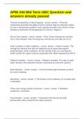
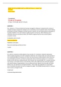
-Sarcomere structure:
-are cylindrical and vary in length between 1 and 5 micrometers depending on the length of the
muscle.
-are comprised of several interlacing’ ‘thick'' and ''thin'' filaments, which slide past each other as the
sarcomere changes length.
-thick filaments are formed by hundreds of myosin tails, thin filaments are composed of three
proteins: actin, tropomyosin, and troponin.
actin: provides binding sites for the myosin heads, the binding sites are spaced at regular intervals
along the thin filament.
tropomyosin and troponin: help to regulate force production by exposing these binding sites only
when the muscle is activated by the nervous system and in presence of calcium.
titin: attaches each thick filament to the ends of the sarcomere (which we call Z-lines or Z-discs), plays
an important role in passive force generation.
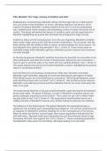
-sarcomeres are arranged in series and parallel within skeletal muscle in a regular pattern, resulting in
striations, alternating light and dark bands viewed under a microscope.
-Z-discs and M-discs are also called lines because that's how they look like in 2D.
- A bands (dark) appear in regions containing myosin and the I bands (light band) consist of the thin
filaments of 2 sarcomeres.
-the thin actin filaments are anchored at one end to the Z-discs.
-the thick myosin filaments attach at one end to structures in the M-discs and at the other end are
tethered to the Z-discs by titin molecules.
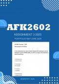
Nebulin: span from the Z line to the actin filaments, acts as a template for the thin filament assembly.
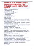
T tubule: are extensions of the cell membrane (sarcolemma) that associates with the terminal cisternae
, 1.Microscopic anatomy: sarcomeric anatomy, myofibrils:
Muscle consists of fascicules, fascicules consist of muscle fibers, and muscle fibers consist of myofibrils
and myofibrils consist of sarcomeres.
-Sarcomere structure:
-are cylindrical and vary in length between 1 and 5 micrometers depending on the length of the
muscle.
-are comprised of several interlacing’ ‘thick'' and ''thin'' filaments, which slide past each other as the
sarcomere changes length.
-thick filaments are formed by hundreds of myosin tails, thin filaments are composed of three
proteins: actin, tropomyosin, and troponin.
actin: provides binding sites for the myosin heads, the binding sites are spaced at regular intervals
along the thin filament.
tropomyosin and troponin: help to regulate force production by exposing these binding sites only
when the muscle is activated by the nervous system and in presence of calcium.
titin: attaches each thick filament to the ends of the sarcomere (which we call Z-lines or Z-discs), plays
an important role in passive force generation.
-sarcomeres are arranged in series and parallel within skeletal muscle in a regular pattern, resulting in
striations, alternating light and dark bands viewed under a microscope.
-Z-discs and M-discs are also called lines because that's how they look like in 2D.
- A bands (dark) appear in regions containing myosin and the I bands (light band) consist of the thin
filaments of 2 sarcomeres.
-the thin actin filaments are anchored at one end to the Z-discs.
-the thick myosin filaments attach at one end to structures in the M-discs and at the other end are
tethered to the Z-discs by titin molecules.
Nebulin: span from the Z line to the actin filaments, acts as a template for the thin filament assembly.
T tubule: are extensions of the cell membrane (sarcolemma) that associates with the terminal cisternae